Bacterial community trends associated with sea scallop, Placopecten magellanicus, larvae in a hatchery system.

Scallops are a diverse animal group of marine bivalve mollusks (family Pectinidae) with global distribution in coastal waters, and Atlantic deep-sea scallops, Placopecten magellanicus, are found along the eastern coast of the United States and Canada. Scallops’ reproductive potential and industry demand make them a prime target for hatchery- and farm-based production, and this has been successfully achieved in bay scallops, but not in sea scallops. Currently, hatcheries collect wild sea scallop adults, or maintain cultured broodstocks, and spawn them in their facilities with the intention of forming a plentiful population to grow to adulthood, spawn, and sell to create a sustainable production cycle while also reducing disruption to the scallops’ natural habitat.

Unfortunately, in sea scallop hatcheries the last two weeks of the larval maturation phase, the veliger-stage, is plagued by large mortality events, going from 60 million sea scallop larvae down to several thousand individuals in a span of 48 hours. Survival of clutches to maturity remains very low, with an industry-standard rate around 1%. This drastic winnowing of larvae reduces the availability of cultured sea scallop spat for farmers, forcing sea scallop farms to rely almost exclusively on sea scallop spat collected from wild populations for stock and is seen as a bottleneck for growth of the industry and achieving sustainable harvests. Hatchery larval die-off is well-demonstrated not to be caused by inadequate diet, lighting, temperature, or atmospheric pressure in aquaculture facilities compared to wild conditions.

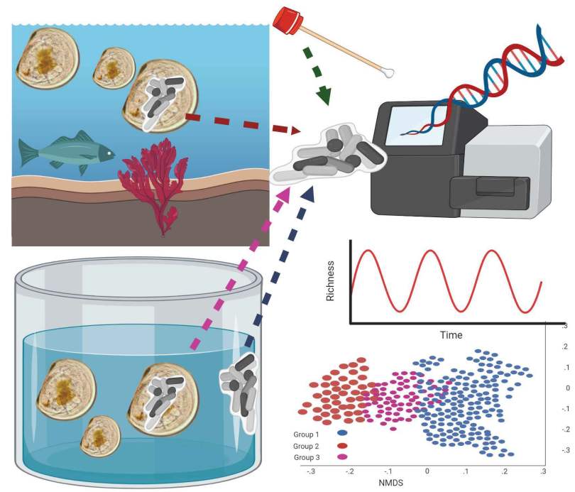
This project wanted to know if there was any clue in the bacteria that associate with larvae, or with the tanks they are in. In particular, hactcheries are worried about certain species of bacteria in the genus Vibrio, as they can cause disease to scallops and/or people, but it is tricky to study them because there are many species which do nothing at all. This project is part of another experiment to examine some of the Vibrio we found in tanks.

We sampled from some wild larvae, hatchery larvae, and from tank biofilms to indentify what was there. There were two styles of tank setup, and we collected from used tanks as well as tanks after they had been cleaned and refilled with filtered seawater.

One of the surprising things we found, was that the bacterial communities in biofilms along the sides of larvae tank were more similar to each other (clustering) when samples were collected during the same phase of the lunar cycle. Bacterial richness and community similarity between tank samples fluctuated over the trial in repeated patterns of rise and fall, which showed some correlation to lunar cycle  where richness is high when the moon is about 50% and richness is low during new and full moon phases. This may be a proxy for the effects of spring tides and trends in seawater bacteria and phages which are propagated into hatchery tanks. The number of days since the full moon was significantly correlated with bacterial community richness in tanks: low during the full moon, peaking ~ 21 days after the full moon, and decreasing again at the next full moon.  

Fig. 7. Constrained ordination of bacterial communities in tank samples. Each point represents the bacterial community from one sample. Similarity between samples was calculated using Distance-based Redundancy Analysis (dbDRA), and significant model factors (anova, p < 0.01) are displayed with arrow lengths relative to their importance in the model (f value). The shape of points indicates whether swabbing was either immediately after filtered seawater has been used to fill the tank (cleaned, refilled) or 48 hours after (dirty, drained). Tank setup indicates if water was static, constantly filtered and recirculated in a flow-through system, or setup information was not available (n/a).

These results along with future work, will inform hatcheries on methods that will increase larval survival in these facilities, for example, implementing additional filtering or avoiding seawater collection during spring tides, to reduce certain bacterial taxa of concern or promoting a more diverse microbial community which would compete against pathogens.

Bacterial community trends associated with sea scallop, Placopecten magellanicus, larvae in a hatchery system.

Authors: Suzanne L. Ishaq1*, Sarah Hosler1, Adwoa Dankwa1, Phoebe Jekielek2, Damian C. Brady3, Erin Grey4,5, Hannah Haskell6, Rachel Lasley-Rasher6, Kyle Pepperman7, Jennifer Perry1, Brian Beal8, Timothy J. Bowden1

Affiliations:1 School of Food & Agriculture, University of Maine, Orono ME 044692 Ecology and Environmental Sciences, University of Maine, Orono ME 044733 School of Marine Sciences, Darling Marine Center, University of Maine, Walpole ME 045734 School of Biology and Ecology, University of Maine, Orono ME 044695 Maine Center for Genetics in the Environment, University of Maine, Orono ME 044696 Department of Biological Sciences, University of Southern Maine, Portland ME 041037 Downeast Institute, Beals, ME 046118 Division of Environmental & Biological Sciences, University of Maine at Machias, Machias, ME 04654

Abstract

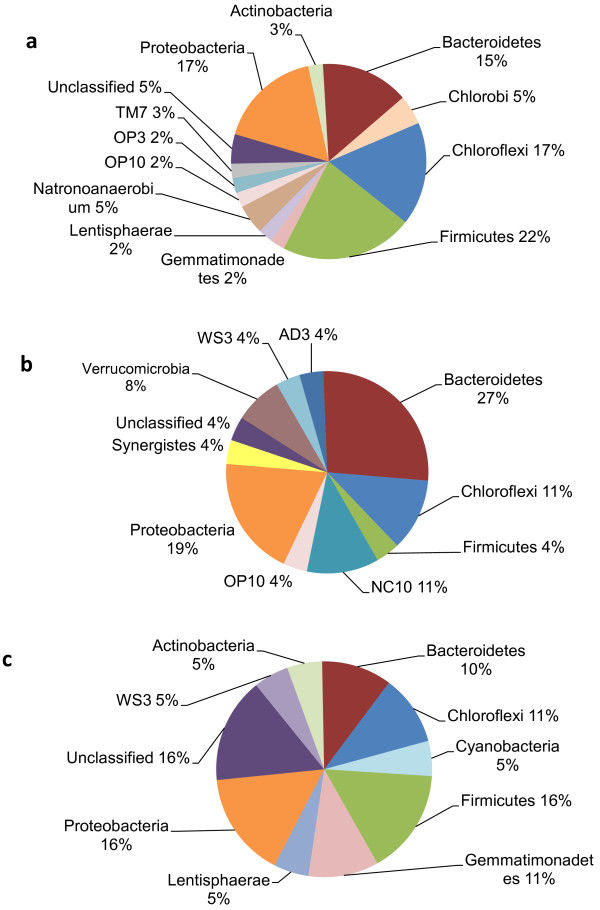
Atlantic sea scallops, Placopecten magellanicus, are the most economically important marine bivalves along the northeastern coast of North America. Wild harvest landings generate hundreds of millions of dollars, and wild-caught adults and juvenile spat are increasingly being cultured in aquaculture facilities and coastal farms. However, the last two weeks of the larval maturation phase in hatcheries are often plagued by large mortality events. Research into other scallop- and aquacultured-species point to bacterial infections or altered functionality of microbial communities which associate with the host. Despite intense filtering and sterilization of seawater, and changing tank water every 48 hours, harmful microbes can still persist in biofilms and mortality is still high. There are no previous studies of the bacterial communities associated with the biofilms growing in scallop hatchery tanks, nor studies with wild or hatchery sea scallops. We characterized the bacterial communities in veliger-stage wild or hatchery larvae, and tank biofilms using the 16S rDNA gene V3-V4 region sequenced on the Illumina MiSeq platform. Hatchery larvae had lower bacterial richness (number of bacteria taxa present) than the wild larvae and tank biofilms, and hatchery larvae had a similar bacterial community (which taxa were present) to both wild larvae and tank biofilms. Bacterial richness and community similarity between tank samples fluctuated over the trial in repeated patterns of rise and fall, which showed some correlation to lunar cycle that may be a proxy for the effects of spring tides and trends in seawater bacteria and phages which are propagated into hatchery tanks. These results along with future work, will inform hatcheries on methods that will increase larval survival in these facilities, for example, implementing additional filtering or avoiding seawater collection during spring tides, to reduce bacterial taxa of concern or promote a more diverse microbial community which would compete against pathogens.

Acknowledgements

The authors would like to thank the staff at the Downeast Institute for supporting the development and implementation of this project, as well as for financially supporting the DNA sequencing; Meredith White of Mook Sea Farm for sharing her expertise and collecting biofilm samples; the Darling Marine Center for sharing their expertise and collecting biofilm samples; and the Sea Scallop Hatchery Implementation (Hit) Team for their expertise, review of this work, and funding support, who are financially supported by the Atlantic States Marine Fisheries Commission and Michael & Alison Bonney. The authors thank Lilian Nowak for assistance with related lab work to this project, and the Map Top Scholars Program for related financial support. The authors also thank Nate Perry for helping us collect wild scallop larvae. All authors have read and approved the final manuscript. This project was supported by the USDA National Institute of Food and Agriculture through the Maine Agricultural & Forest Experiment Station, Hatch Project Numbers: ME0-22102 (Ishaq), ME0-22309 (Bowden), and ME0-21915 (Perry); as well through NSF #OIA-1849227 to Maine EPSCoR at the University of Maine (Grey). This project was supported by an Integrated Research and Extension Grant from the Maine Food and Agriculture Center, with funding from the Maine Economic Improvement Fund.

Ishaq, S.L., Hosler2, S., Dankwa, A., Jekielek, P., Brady, D.C., Grey, E., Haskell, H., Lasley-Rasher, R., Pepperman, K., Perry, J., Beal, B., Bowden, T.J. 2023. Bacterial community trends associated with sea scallop, Placopecten magellanicus, larvae in a hatchery system. Aquaculture Reports 32: 101693.

Warmer water temperature and epizootic shell disease reduces diversity but increases cultivability of bacteria on the shells of American Lobster (Homarus americanus).

This collaborative paper investigates what happens to bacterial communities on healthy and sick lobsters as they experience different water temperatures for a year: “Water temperature and disease alters bacterial diversity and cultivability from American Lobster (Homarus americanus) shells.”

Woman sitting outside.

I joined this project back in the summer of 2020, towards the end of my first year at UMaine, when I was given a large 16S rRNA gene sequence dataset of bacterial communities from the shells of lobsters. I had been asking around for data as a training opportunity for Grace Lee, who at the time was an undergraduate at Bowdoin College participating in the abruptly cancelled summer Research Experience for Undergrads program at UMaine in summer 2020. Instead, Grace joined my lab as a remote research assistant and we worked through the data analysis over the summer and fall. Grace has since graduated with her Bachelor’s of Science in Neuroscience, obtained a Master’s of Science at Bowdoin, and is currently a researcher at Boston Children’s Hospital while she is applying to medical school.

My first point of contact on the project was Jean MacRae, an Associate Professor of Civil and Environmental Engineering at UMaine, who was the one to lend me the data and who had been working on bacterial community sequencing on other projects which I’ve been involved in. Jean has been involved with MSE, and this is our fourth publication together making her the collaborator at UMaine I have co-authored with the most (although it is a tight race 🙂 ).

Four professors wearing full regalia, as well as face masks, posing for a photo in a hallway.

Jean introduced me to the original research team, including Debbie Bouchard, who is the Director of the Aquaculture Research Institute and was researching epizootic shell disease in lobsters for her PhD dissertation several years ago; Heather Hamlin, Professor and Director of the School of Marine Sciences; Scarlett Tudor (not pictured), the Education and Outreach Coordinator at the ARI; and Sarah Turner (not pictured), Scientific Research Specialist at ARI. The ARI team is involved in a lot of large-scale aquaculture research, education, and outreach to the industry here in Maine, and the collaborative work I have been doing with them has been a new an engaging avenue of scientific study for me.

In 2022, the research team, along with social science Masters student Joelle Kilchenmann, published a perspective/hypothesis piece which explored unanswered questions about how the movement of microbes, lobsters, and climate could affect the spread of epizootic shell disease in lobsters off the coast of Maine. That perspective paper was a fun exercise in hypothesis generation and asking ‘what if‘?

A steamed lobster on a plate.

This paper is more grounded, and features work that was started in 2016. It examines bacterial communities on the shells of lobsters which were captured off the coast of Southern Maine and maintained in aquarium tanks for over a year. The lobsters were split into three treatment groups: those which were kept in water temperatures that mimicked what they would experience in Southern Maine, colder water to simulated what they would experience in Northern Maine, and hotter water to simulate what they would experience in Southern New England over that year. The original project team wanted to know if temperatures would make a different to their health or microbial communities.

Figure S8. Water temperature regimes, related to STAR Methods. A. Temperatures were obtained through the National Oceanographic Data Center (NODC). NODC temperatures reflect those recorded near Eastport, ME (A); Portland, ME (B); and an average of temperatures from Woods Hole, MA (C) and New Haven, CT (D) was used to represent Southern New England. B. Annual temperature cycles used in this project to represent Southern New England (SNE), Southern Maine (SME) and Northern Maine (NME).

The original project team swabbed lobster shells to obtain bacteria to try and grow in the lab, as well as DNA to sequence and identify whole bacterial communities. Grace and I performed the data analysis to identify which taxa were present in those communities, what happened over time or when the water temperature changed, and what bacteria were present or not in lobsters which died during the study.

Figure S11. Lobster carapace sampling using a sterile cotton swab to obtain bacterial communities from the shell surface, related to STAR Methods. The right side of the dorsolateral area of the cephalothorax was sampled for the baseline sampling, the left side for the Time 1, and the right side again for Time 2.

In addition to wanting to know about temperature, we wanted to know specifically how temperature would affect the bacteria if the lobsters had epizootic shell disease. It is not known what causes epizootic shell disease (which is why it is called ‘epizootic’), but it manifests as pitting in the shells of lobsters. Over time, the pitting can weaken shells and make it difficult for the lobster to molt, or make the lobster susceptible to predators or microbial infections. This type of shell disease had been a huge problem in Southern New England over the past few decades, and in Maine we have seen more cases over time.

Four panels of lobsters showing the progression of a healthy lobster to ones with more and more pitting in their shells.
Figure S10. Examples of lobster shell disease indices, related to STAR Methods. A) 0, no observable signs of disease, B) 1+, shell disease signs on 1-10% of the shell surface, C) 2+, shell disease signs on 11-50% of the shell surface, D) 3+, shell disease signs on > 50% of the shell surface.

The highlights of this project are here, but you can click the link below to read the entire study and what happened to lobster health and lobster microbes over time.

  • Shell bacteria from healthy lobsters, often overlooked, were included in the study.
  • Hotter and colder water temperatures affected shell bacterial communities.
  • Epizootic shell disease reduced bacterial diversity on lobster shells.
  • Epizootic shell disease could be induced or exacerbated by the loss of commensal bacteria from shells.

“Water temperature and disease alters bacterial diversity and cultivability from American Lobster (Homarus americanus) shells.”

Suzanne L. Ishaq1,2,, Sarah M. Turner2,3, Grace Lee4,5,M. Scarlett Tudor2,3, Jean D. MacRae6, Heather Hamlin2,7, Deborah Bouchard2,3

  • 1 School of Food and Agriculture; University of Maine; Orono, Maine, 04469; USA.
  • 2 Aquaculture Research Institute; University of Maine; Orono, Maine, 04469; USA.
  • 3 Cooperative Extension; University of Maine; Orono, Maine, 04469; USA.
  • 4 Department of Neuroscience, Bowdoin College, Brunswick, ME 04011; USA.
  • 5 Boston Children’s Hospital, Boston, MA 02115; USA.
  • 6 Department of Civil and Environmental Engineering; University of Maine; Orono, Maine, 04469; USA.
  • 7 School of Marine Sciences; University of Maine; Orono, Maine, 04469; USA.

iScience 26(5): 106606.

Summary

The American lobster, Homarus americanus, is an economically valuable and ecologically important crustacean along the North Atlantic coast of North America. Populations in southern locations have declined in recent decades due to increasing ocean temperatures and disease, and these circumstances are progressing northward. We monitored 57 adult female lobsters, healthy and shell-diseased, under three seasonal temperature cycles for a year, to track shell bacterial communities using culturing and 16S rRNA gene sequencing, progression of ESD using visual assessment, and antimicrobial activity of hemolymph. The richness of bacterial taxa present, evenness of abundance, and community similarity between lobsters was affected by water temperature at the time of sampling, water temperature over time based on seasonal temperature regimes, shell disease severity, and molt stage. Several bacteria were prevalent on healthy lobster shells but missing or less abundant on diseased shells, although some bacteria were found on all shells regardless of health status.

Many questions remain unanswered about the role of microbial transmission in epizootic shell disease in American lobsters (Homarus americanus).

Lobsters are an iconic part of Maine culture, from cuisine to interior decorating to way of life. The Gulf of Maine boasts large lobster landings every year, but as the waters here continue to warm at a faster rate than other nearby coastal regions, there are concerns that this boon might eventually pass us by as lobsters migrate further north in search of colder waters. In addition to rising temperatures along the northeastern coast, we’ve seen an increase in epizootic shell disease (ESD) in the last few decades. ESD causes degradation and pitting of the lobster shell which can leave them susceptible to predation or to harsh weather conditions. There are bacteria living on the shell of healthy lobsters, and it’s not clear how they are involved in ESD in the wild because it is difficult to replicate this disease in an aquaculture facility.

In this perspective piece, we consider how shell microbes might be involved. Marine environments have a thriving microbial community which can change rapidly when currents, storms, filter feeders, or viruses which target microbes roll through. Some of these water or soil microbes end up on lobster shells, and water currents can also lift microbes off and move them elsewhere. The other authors and I wanted to highlight some of these possibilities and what we still don’t know about lobster microbes and health.

A steamed lobster on a plate.

This perspective piece is part of a larger, collaborative project on lobster shell disease and warming ocean waters was begun by researchers at the Aquaculture Research Institute: Debbie Bouchard, Heather Hamlin, Jean MacRae, Scarlett Tudor, and later Sarah Turner as a grad student. I was invited to participate in the data analysis aspect two years ago.

At the time, Grace Lee was a rising senior at Bowdoin College, and accepted to my lab for the UMaine REU summer 2020 session, which was canceled. Instead, I hired Grace to perform DNA sequence analysis remotely, by independently learning data analysis following the teaching materials I had generated for my sequencing class.  I invited Joelle Kilchenmann to this piece after a series of conversations about microbes and social equity, because her graduate work in Joshua Stoll’s lab focuses on lobster fishing communities in Maine and understanding the challenges they face.


Ishaq, S.L., Turner, S.M., Tudor, M.S.,  MacRae, J.D., Hamlin, H., Kilchenmann, J., Lee1, G., Bouchard, D. 2022. Many questions remain unanswered about the role of microbial transmission in epizootic shell disease in American lobsters (Homarus americanus). Frontiers in Microbiology 13: 824950.

This was an invited contribution to a special collection: The Role of Dispersal and Transmission in Structuring Microbial Communities

Abstract: Despite decades of research on lobster species’ biology, ecology, and microbiology, there are still unresolved questions about the microbial communities which associate in or on lobsters under healthy or diseased states, microbial acquisition, as well as microbial transmission between lobsters and between lobsters and their environment. There is an untapped opportunity for metagenomics, metatranscriptomics, and metabolomics to be added to the existing wealth of knowledge to more precisely track disease transmission, etiology, and host-microbe dynamics. Moreover, we need to gain this knowledge of wild lobster microbiomes before climate change alters environmental and host-microbial communities more than it likely already has, throwing a socioeconomically critical industry into disarray. As with so many animal species, the effects of climate change often manifests as changes in movement, and in this perspective piece, we consider the movement of the American lobster (Homarus americanus), Atlantic ocean currents, and the microorganisms associated with either.


Related presentations

Ishaq*, S.L., Lee, G., MacRae, J., Hamlin, H., Bouchard, D. “The effect of simulated warming ocean temperatures on the bacterial communities on the shells of healthy and epizootic shell diseased American Lobster (Homarus americanus).” Ecological Society of America 2021. (virtual). Aug 2-6, 2021. (accepted talk)

Ishaq*, S.L., Lee, G., MacRae, J., Hamlin, H., Bouchard, D. The Effect Of Simulated Warming Ocean Temperatures On The Bacterial Communities On The Shells Of Healthy And Epizootic Shell Diseased American Lobster (Homarus americanus)ASM Microbe/ISME World Microbe Forum 2021 (virtual). June 20-24, 2021. (poster)

Determination of the microbial community in the rumen and fecal matter of lactating dairy cows fed on reduced-fat dried distillers grains with solubles.

Ruminants, like sheep, goats, cows, deer, moose, etc.,  have a four-chambered stomach, the largest of which is called the rumen.  The rumen houses symbiotic microorganisms which break down plant fibers that the animal can’t digest on its own.  It’s estimated that up to 80% of a ruminant’s energy need is met from the volatile fatty acids (also called short-chain fatty acids) that bacteria produce from digesting fiber, and that up to 85% of a ruminant’s protein need is met from microbial proteins.

A lot of factors can be manipulated to help get the most out of one’s diet, including the source and processing method of the ingredients – in most cases in livestock feed: plants. Growing plants for animal feed can be expensive, and often nutrients in plants become more available to the animal after the plant has been processed/broken down in some way. This sometimes allows for food byproducts to be reused for animal feed, and one common example is used brewers’ grains. Once the grains have been fermented to produce alcohol, the simple sugars have been used up but a lot of the complex sugar carbohydrates – in other words: fiber – are left over. Ruminants don’t need simple sugars, but they do need a lot of fiber, and brewers’ grains have been investigated for their usefulness for animal nutrition because they are a cheap, readily-available, and common source of fiber, as well as protein.

The original experiment for this work took place several years ago, and involved an animal feeding trial which added reduced-fat distillers’ grains with solubles into dairy cattle feed. The research team found no negatives effect on milk production or animal health, and that work was previously published. To add to that project, the original research team wanted to know if the diet would drastically change the bacterial community living in the rumen, which would have implications for feed digestion and animal health.

A collaborator of mine donated the cow microbial community DNA data to my AVS 590 special topics in DNA Sequencing Data Analysis course in spring 2020 (now formally registered as AVS 454/554). I worked with UMaine graduate students Adwoa Dankwa and Usha Humagain over the semester to train them in coding and develop the manuscript. The diet only had minimal effects on the bacterial community profiles, which in this case is a good finding – we want to be able to feed a cheap, nutritional source like distillers’ grains without harming the cow or its microbes.


Dankwa, A.S., U. Humagain, S.L. Ishaq, C.J. Yeoman, S. Clark , D.C. Beitz, and E. D. Testroet. 2021. Determination of the microbial community in the rumen and fecal matter of lactating dairy cows fed on reduced-fat dried distillers grains with solubles. Animal 15(7):100281.

Abstract

Reduced-fat dried distillers’ grains with solubles (RF-DDGS) is a co-product of ethanol production and contains less fat than traditional distillers’ grains. The fat in corn is ~ 91% unsaturated, and it is toxic to rumen microorganisms so it could influence the composition of the rumen microbiome. It has been demonstrated that RF-DDGS is a suitable ration ingredient to support the high-producing dairy cow, and this feedstuff is a promising alternative protein source for lactating dairy cows. The current study aims to better understand the effect of RF-DDGS on the rumen and fecal bacterial composition in lactating dairy cows. Thirty-six multiparous (2 or 3), mid-lactation Holstein cows (BW = 680 ± 11 kg; 106 ± 27 DIM) were randomly assigned to two groups which were fed a control diet made up of corn, corn silage, and alfalfa hay supplemented with expeller soybean meal or with added RF-DDGS (20% of the dry matter (DM)) containing approximately 6.0% fat. Whole rumen contents (rumen fluid and digesta; esophageal tubing method) and feces (free catch method) were collected on day 35 of the experimental period, after the 14-d acclimation period. Rumen contents and feces from each cow were used for DNA extraction. The bacterial community composition in rumen and fecal samples was assessed via the 16S rRNA gene by using the Illumina MiSeq sequencing platform. Bacteroidetes, Actinobacteria, and Firmicutes were the most abundant phyla in rumen contents. The fecal microbiota was dominated by the phyla Firmicutes and Bacteroidetes, as well as Actinobacteria and Chloroflexi. RF-DGGS increased bacterial richness, evenness, and Shannon diversity in both rumen and fecal samples and was associated with several taxa that had different abundance in treatment versus control comparisons.  The RF-DGGS, however, did not significantly alter the bacterial community in the rumen or feces. In general, these findings demonstrated that dietary inclusion of RF-DDGS did not impose any serious short-term (within 30 days) health or production consequences, as would be expected. With this study, we present further evidence that inclusion of 20% (DM basis) RF-DDGS in the diet of lactating dairy cows can be done without consequence on the microbiome of the rumen.

Implications

Reduced-fat dried distillers’ grains with solubles is a quality, economical, and readily available protein source demonstrated to support the protein needs of high-producing dairy cows. In this study, the rumen and fecal bacterial communities of lactating dairy cows were not significantly influenced by 20% (dry matter basis) reduced-fat dried distillers’ grains with solubles and did not impose serious short-term (within 30 days) health or production consequences. This diet could potentially be introduced into Total Mixed Ration feeding of dairy cattle given the fact that it is readily available and relatively economical.

Bacterial transfer from Pristionchus entomophagus nematodes to the invasive ant Myrmica rubra and the potential for colony mortality in coastal Maine.

A collaborative paper on bacterial transfer in insects and the possible ecological impacts of that in the wild has been published in iScience! This work began a decade ago in the labs of Dr. Ellie Groden, recently retired Professor of Entomology in the School of Biology and Ecology at the University of Maine, and later Dr. Patricia Stock, a Professor in the School of Animal and Comparative Biomedical Sciences at the University of Arizona, who were investigating colony collapse of European fire ants (Myrmica rubra) which are invasive to Maine. The ants have a nasty bite, and can dramatically disturb the local plant and insect wildlife in coastal Maine.

Slide from Ishaq et al. Entomology 2020 presentation

When these invasive ant colonies collapsed, Drs. Groden and Stock wanted to find out why, as a possible means of developing a biological control strategy. It was thought that particular nematodes would ingest soil bacteria, and transfer it to ants once the worms invaded ant tissues to complete parts of their life cycle. This particular worm infection doesn’t kill the ants, but perhaps the soil bacteria were. Ants were collected from different colony sites, and investigations on the nematode worms inhabiting the ants were conducted.

Slide from Ishaq et al. Entomology 2020 presentation

Most of the work for this project was completed several years ago, with the exception of DNA sequencing data from a bacterial transfer experiment. I was added to the project by my collaborator at UMaine, Dr. Jean MacRae, an Associate Professor in the Department of Civil and Environmental Engineering who introduced me to the research team and shared the 16S rRNA dataset to use in my AVS 590 data analysis class in spring 2020. That semester was when the pandemic hit, and forced the course to move to remote-only instruction in March. UMaine graduate students Alice Hotopp and Sam Silverbrand were taking the class and learning 16S analysis on this dataset, and I mentored them through the analysis all the way to manuscript writing despite the incredible challenges that spring threw our way.

At the completion of the course, we shared the draft manuscript with the rest of the research team, who mentioned that several undergraduate honor’s theses had been written about the earlier experiment, but never published in a scientific journal. The team spent summer 2020 combining the three papers into one massive draft. The pandemic slowed down manuscript review, understandably, but I’m pleased to say that it was accepted for publication! In addition, this collaboration has led to further collaborations in the Ishaq Lab, several presentations (listed below), and is Sam’s first scientific publication, congrats Sam!!

Alice Hotopp, A., Samantha Silverbrand, Suzanne L. Ishaq, Jean MacRae, S. Patricia Stock, Eleanor Groden. “Can a necromenic nematode serve as a biological Trojan horse for an invasive ant?Ecological Society of America 2021 (virtual). Aug 2-6, 2021 (accepted poster).

Ishaq*, S.L., Hotopp, A., Silverbrand, S.,   MacRae, J.,  Stock, S.P.,  Groden, E. “Can a necromenic nematode serve as a biological Trojan horse for an invasive ant?” Entomological Society of America 2020 (virtual). Nov 15-25, 2020. (invited talk)

Press/Interviews

It began on an anthill in Maine“, Sue Ishaq and Ellie Groden, iScience Backstories, Dec 17, 2021.

UMaine researchers want to use nematodes to kill fire ants.”, Julia Bayly, Bangor Daily News, July 29, 2021.

Bacteria from nematodes could be used to kill fire ants, UMaine research reveals”, Marcus Wolf, University of Maine news, July 27, 2021.

Illustrated image of a cross section of the ground. A light brown ant is pictured in the ground along with a microbe. Text to the left of the image reads, "Can a necromenic nematode serve as a biological Trojan horse for an invasive ant?". The names of six professors are listed below the text and image at the bottom left. In the bottom right corner, text reads, "The University of Maine" with "The University of Arizona" below it.

IshaqS.L., A. Hotopp2, S. Silverbrand2, J.E. Dumont, A. Michaud, J. MacRae, S. P. Stock, E. Groden. 2021. Bacterial transfer from Pristionchus entomophagus nematodes to the invasive ant Myrmica rubra and the potential for colony mortality in coastal Maine. iScience. In Press. Impact 5.08.

Abstract

The necromenic nematode Pristionchus entomophagus has been frequently found in nests of the invasive European ant Myrmica rubra in coastal Maine, United States, and may contribute to ant mortality and collapse of colonies by transferring environmental bacteria. Paenibacillus and several other bacterial species were found in the digestive tracts of nematodes harvested from collapsed ant colonies. Serratia marcescens, Serratia nematodiphila, and Pseudomonas fluorescens were collected from the hemolymph of nematode-infected wax moth (Galleria mellonella) larvae.

Virulence against waxworms varied by site of origin of the nematodes. In adult nematodes, bacteria were highly concentrated in the digestive tract with none observed on the cuticle. In contrast juveniles had more on the cuticle than in the digestive tract. .  Host species was the primary factor affecting bacterial community profiles, but Spiroplasma sp. and Serratia marcescens sequences were shared across ants, nematodes, and nematode-exposed G. mellonella larvae. 

Pelleted-hay alfalfa feed increases sheep wether weight gain and rumen bacterial richness over loose-hay alfalfa feed.

Ruminants, like sheep, goats, cows, deer, moose, etc.,  have a four-chambered stomach, the largest of which is called the rumen.  The rumen houses symbiotic microorganisms which break down plant fibers that the animal can’t digest on its own.  It’s estimated that up to 80% of a ruminant’s energy need is met from the volatile fatty acids (also called short-chain fatty acids) that bacteria produce from digesting fiber, and that up to 85% of a ruminant’s protein need is met from microbial proteins.

A lot of factors can be manipulated to help get the most out of one’s diet, including adjusting ingredients for water content, palatability, ease of chewing, and how easy the ingredients are to digest.  For example, highly fibrous foods with larger particles/pieces require more chewing, as well as a longer time spent in the rumen digesting so that microorganisms have plenty of time to break the chemical bonds of large molecules.  Smaller food particles can reduce the time and effort spent chewing, allow for more surface area on plant fibers for microorganisms to attach to and digest faster, and speed up the movement of food through the digestive tract.  On the other hand, moving food too quickly could reduce the amount of time microorganisms can spend digesting, or time the ruminant can absorb nutrients across their GI tract lumen, or cause slow-growing microbial species to wash out.

Surprisingly, almost no work has investigated the effect of diet particle size on the community, despite knowing that microbial digestion is contingent on the ability to attach to and process complex nutrient structures.  In this study, we observed the effect of particle size on rumen bacteria, by feeding long-stem (loose) alfalfa hay compared to a ground and pelleted version of the same alfalfa in yearling sheep wethers. 

The pelleted-hay diet group had a greater increase in bacterial richness, including common fibrolytic rumen inhabitants, which may explain the increase in average daily gain and feed efficiency in this group.

Fig 2. Observed bacterial richness (A) and Shannon diversity (B) in the rumen of wethers on loose-hay or pelleted-hay alfalfa diets. Significance was determined at p < 0.05, by linear mixed model for observed SVs and Conover test for Shannon diversity, with sheep ID as a fixed effect.
Fig 5. Discriminatory rumen bacterial sequence variance by treatment group for wethers receiving loose-hay or pelleted-hay alfalfa diet treatments.Significance (p < 0.05) determined by binomial test. 

Ishaq SL, Lachman MM, Wenner BA, Baeza A, Butler M, Gates E, et al. (2019) Pelleted-hay alfalfa feed increases sheep wether weight gain and rumen bacterial richness over loose-hay alfalfa feed. PLoS ONE 14(6): e0215797. Article.

Abstract

Diet composed of smaller particles can improve feed intake, digestibility, and animal growth or health, but in ruminant species can reduce rumination and buffering–the loss of which may inhibit fermentation and digestibility. However, the explicit effect of particle size on the rumen microbiota remains untested, despite their crucial role in digestion. We evaluated the effects of reduced particle size on rumen microbiota by feeding long-stem (loose) alfalfa hay compared to a ground and pelleted version of the same alfalfa in yearling sheep wethers during a two-week experimental period. In situ digestibility of the pelleted diet was greater at 48 h compared with loose hay; however, distribution of residual fecal particle sizes in sheep did not differ between the dietary treatments at any time point (day 7 or 14). Both average daily gain and feed efficiency were greater for the wethers consuming the pelleted diet. Observed bacterial richness was very low at the end of the adaptation period and increased over the course of the study, suggesting the rumen bacterial community was still in flux after two weeks of adaptation. The pelleted-hay diet group had a greater increase in bacterial richness, including common fibrolytic rumen inhabitants. The pelleted diet was positively associated with several Succiniclasticum, a Prevotella, and uncultured taxa in the Ruminococcaceae and Rickenellaceae families and Bacteroidales order. Pelleting an alfalfa hay diet for sheep does shift the rumen microbiome, though the interplay of diet particle size, retention and gastrointestinal transit time, microbial fermentative and hydrolytic activity, and host growth or health is still largely unexplored.

Feature image credit: Pellet Mill

Zinc amino acid supplementation alters yearling ram rumen bacterial communities but zinc sulfate supplementation does not.

Zinc is an important mineral in your diet; it’s required by many of your enzymes and having too much or too little can cause health problems. We know quite a bit about how important zinc is to sheep, in particular for their growth, immune system, and fertility.  We also know that organically- versus inorganically-sourced zinc differs in its bio-availability, or how easy it is for cells to access and use it.  Surprisingly, we know nothing about how different zinc formulations might affect gut microbiota, despite the knowledge that microorganisms may also need zinc.

This collaborative study was led by Dr. Whit Stewart and his then-graduate student, Chad Page, while they were at Montana State University (they are now both at the University of Wyoming).   Chad’s work focused on how different sources of zinc affected sheep growth and performance (previously presented, publication forthcoming), and I put together this  companion paper examining the effects on rumen bacteria. Unfortunately, the article is not currently open-access.


Ishaq, S.L., Page, C.M., Yeoman, C.J., Murphy, T.W., Van Emon, M.L., Stewart, W.C. 2019. Zinc amino acid supplementation alters yearling ram rumen bacterial communities but zinc sulfate supplementation does not. Journal of Animal Science 97(2):687–697. Article.

Abstract

Despite the body of research into Zn for human and animal health and productivity, very little work has been done to discern whether this benefit is exerted solely on the host organism, or whether there is some effect of dietary Zn upon the gastrointestinal microbiota, particularly in ruminants. We hypothesized that 1) supplementation with Zn would alter the rumen bacterial community in yearling rams, but that 2) supplementation with either inorganically-sourced ZnSO4, or a chelated Zn amino acid complex, which was more bioavailable, would affect the rumen bacterial community differently. Sixteen purebred Targhee yearling rams were utilized in an 84 d completely-randomized design, and allocated to one of three pelleted dietary treatments: control diet without fortified Zn (~1 x NRC), a diet fortified with a Zn amino acid complex (~2 x NRC), and a diet fortified with ZnSO4 (~2 x NRC). Rumen bacterial community was assessed using Illumina MiSeq of the V4-V6 region of the 16S rRNA gene. One hundred and eleven OTUs were found with > 1% abundance across all samples. The genera PrevotellaSolobacteriumRuminococcusButyrivibrioOlsenellaAtopobium, and the candidate genus Saccharimonas were abundant in all samples. Total rumen bacterial evenness and diversity in rams were reduced by supplementation with a Zn-amino-acid complex, but not in rams supplemented with an equal concentration of ZnSO4, likely due to differences in bioavailability between organic and inorganically-sourced supplement formulations. A number of bacterial genera were altered by Zn supplementation, but only the phylum Tenericutes was significantly reduced by ZnSO4 supplementation, suggesting that either Zn supplementation formulation could be utilized without causing a high-level shift in the rumen bacterial community which could have negative consequences for digestion and animal health.

Featured Image Source: Wikimedia Commons

An investigation into rumen fungal and protozoal diversity in three rumen fractions, during high-fiber or grain-induced sub-acute ruminal acidosis conditions, with or without active dry yeast supplementation. 

Ruminal acidosis is a condition in which the pH of the rumen is considerably lower than normal, and if severe enough can cause damage to the stomach and localized symptoms, or systemic illness in cows.  Often, these symptoms result from the low pH reducing the ability of microorganisms to ferment fiber, or by killing them outright.  Since the cow can’t break down most of its plant-based diet without these microorganisms, this disruption can cause all sorts of downstream health problems.  Negative health effects can also occur when the pH is somewhat lowered, or is lowered briefly but repeatedly, even if the cow isn’t showing outward clinical symptoms.  This is known as sub-acute ruminal acidosis(SARA), and can also cause serious side effects for cows and an economic loss for producers.

In livestock, acidosis usually occurs when ruminants are abruptly switched to a highly-fermentable diet- something with a lot of grain/starch that causes a dramatic increase in bacterial fermentation and a buildup of lactate in the rumen.  To prevent this, animals are transitioned incrementally from one diet to the next over a period of days or weeks.  Another strategy is to add something to the diet to help buffer rumen pH, such as a probiotic.  One of the most common species used to help treat or prevent acidosis is a yeast; Saccharomyces cerevisiae.

This paper was part of a larger study on S. cerevisiae use in cattle to treat SARA, the effects of which on animal production as well as bacterial diversity and functionality have already been published by an old friend and colleague of mine, Dr. Ousama AlZahal, and several others.

The main driver of fungal diversity was diet; moving from a high-fiber diet to a high-grain diet (Figure 1) triggered a change in available nutrients (more starch, less fiber), and decreased in rumen pH due to the byproducts related to microbial digestion of those nutrients.  Supplementation with active dry yeast only had minimal effect on fungal populations in the rumen, and did not help recover the fungal community found in healthy cows on a high-fiber diet.  Saccharomyces-related sequences all classified as S. cerevisiae, though to multiple strains, but were not found in >1% mean relative abundance in any treatment group or significantly more abundant in any group. Thus, it was unclear if the yeast supplement was actively part of the rumen fungal community.

PowerPoint Presentation
Figure 1. Relative abundance of rumen fungi genera for cows receiving a high fiber (HF) or high grain (HG) diet, with (Y) or without (C) yeast supplementation. Treatments include high-fiber control (HFC), high-fiber yeast (HFY), high-grain control (HGC), and high-grain yeast (HGY).

Similarly, diet was the major driver of protozoal diversity in the rumen (Figure 2), but there was also a small effect of the yeast supplementation.  Taxonomic diversity was also different between the high-fiber control (what the cows were before) and the high-grain yeast-supplemented group, indicating that yeast supplementation did not recover the initial protozoal community which healthy cows had.

PowerPoint Presentation
Figure 2. Relative abundance of rumen protozoal species for cows receiving a high fiber (HF) or high grain (HG) diet, with (Y) or without (C) yeast supplementation. Treatments include high-fiber control (HFC), high-fiber yeast (HFY), high-grain control (HGC), and high-grain yeast (HGY).

Another large difference was seen in the number and type of species found in three different locations within the rumen: those found in rumen fluid, those found attached to plant material (and presumably digesting it), and those found attached or associated with the rumen wall (epimural-associated).  In cows fed the high-grain diets, there were not enough fungi in the rumen fluid to generate enough sequences for comparison, and the high-grain diet tended to reduce the number of different species found in any location.  Fungal species richness was highest in plant-associated fractions, and there was surprisingly high species richness of fungi which were found along the rumen wall.  Protozoal species richness was likewise reduced by a switch to a high-grain diet, and was highest next to the rumen wall.


Ishaq, S.L., AlZahal, O., Walker, N., McBride, B. 2017. An investigation into rumen fungal and protozoal diversity in three rumen fractions, during high-fiber or grain-induced sub-acute ruminal acidosis conditions, with or without active dry yeast supplementation.  Frontiers in Microbiology 8:1943. Article.

Abstract

Sub-acute ruminal acidosis (SARA) is a gastrointestinal functional disorder in livestock characterized by low rumen pH, which reduces rumen function, microbial diversity, host performance, and host immune function. Dietary management is used to prevent SARA, often with yeast supplementation as a pH buffer. Almost nothing is known about the effect of SARA or yeast supplementation on ruminal protozoal and fungal diversity, despite their roles in fiber degradation. Dairy cows were switched from a high-fiber to high-grain diet abruptly to induce SARA, with and without active dry yeast (ADY, Saccharomyces cerevisiae) supplementation, and sampled from the rumen fluid, solids, and epimural fractions to determine microbial diversity using the protozoal 18S rRNA and the fungal ITS1 genes via Illumina MiSeq sequencing. Diet-induced SARA dramatically increased the number and abundance of rare fungal taxa, even in fluid fractions where total reads were very low, and reduced protozoal diversity. SARA selected for more lactic-acid utilizing taxa, and fewer fiber-degrading taxa. ADY treatment increased fungal richness (OTUs) but not diversity (Inverse Simpson, Shannon), but increased protozoal richness and diversity in some fractions. ADY treatment itself significantly (P < 0.05) affected the abundance of numerous fungal genera as seen in the high-fiber diet: Lewia, Neocallimastix, and Phoma were increased, while Alternaria, Candida Orpinomyces, and Piromyces spp. were decreased. Likewise, for protozoa, ADY itself increased Isotricha intestinalis but decreased Entodinium furca spp. Multivariate analyses showed diet type was most significant in driving diversity, followed by yeast treatment, for AMOVA, ANOSIM, and weighted UniFrac. Diet, ADY, and location were all significant factors for fungi (PERMANOVA, P = 0.0001, P = 0.0452, P = 0.0068, Monte Carlo correction, respectively, and location was a significant factor (P = 0.001, Monte Carlo correction) for protozoa. Diet-induced SARA shifts diversity of rumen fungi and protozoa and selects against fiber-degrading species. Supplementation with ADY mitigated this reduction in protozoa, presumptively by triggering microbial diversity shifts (as seen even in the high-fiber diet) that resulted in pH stabilization. ADY did not recover the initial community structure that was seen in pre-SARA conditions.


Ishaq, S.L.*, O. AlZahal, N. Walker, B. McBride. 2017. Modulation of sub-acute ruminal acidosis by active-dry yeast supplementation and its effect on rumen fungal and protozoal populations in liquid, solid, and epimural fractions.  Congress on Gastrointestinal Function, Chicago, IL, April 2017. (accepted talk).


Featured Image Credit: Wikimedia Commons

Biogeographical Differences in the Influence of Maternal Microbial Sources on the Early Successional Development of the Bovine Neonatal Gastrointestinal tract.

Most studies that examine the microbial diversity of the gastrointestinal tract only look at one or two sample sites, usually the mouth, the rumen in ruminant animals, or the feces.  It can be difficult, expensive, invasive, or fatal to get samples from deep inside the intestinal tract; however many studies have pointed out that anatomical location and local environmental factors (like temperature, pH, host cells, nutrient availability, and exposure to UV light) can dramatically change a microbial community.  Thus, the microbes that we find in feces aren’t always what we would find in the stomach or along the intestines.

On top of that, certain microorganisms have been shown to closely associate with or attach to host cells lining the digestive tract, and the diversity of microbes next to host tissues can be different from what’s at the center of the intestines (the digesta).  This large, collaborative project took samples from nine different sites along the digestive tract of calves over the first 21 days of life to determine how body sites differed from each other, how sites changed over time as the calf matured, and how the lumen-associated bacteria would differ from the digesta-associated bacteria.

41598_2018_21440_Fig1_HTML
Figure 1 Mean bacterial diversity at the phylum level for maternal and calf lumen (A) and mucosal (B) samples.

Samples from the mothers were also taken to understand how maternal microbial influence would affect body sites over time.  One of the most interesting finds of the study regarded colostrum, which is the special and highly-nutritious milk produced in the first 48 hours or so after parturition (birth).  Colostrum milk possessed a high diversity of bacteria, and is not sterile as was once assumed.

Capture
Figure 1 (partial) Mean bacterial diversity at the phylum level for maternal and calf lumen (left) and mucosal (right) samples.

Not only that, but the bacterial community in colostrum had an impact on the bacterial community that developed along the calf digestive tract over time.  Calves received two doses of colostrum on the day of birth which had been aseptically collected from their dams and then fed to them, so that calves received milk but not the microbial influence of nursing and coming into contact with the dam.  After those two meals, calves were switched to milk replacer.  Surprisingly, the influence on the bacterial community wasn’t high on day one and then dropped off.  It increased over the first 21 days of life as bacterial communities from the digestive tract became more similar to bacterial communities found in colostrum (shown below).

colostrum
A poor-quality GIF, showing bacterial communities from the calf digestive tract (each other the colored shaped) becoming more similar to maternal colostrum (milk) samples (grey asterisk) over the first 21 days of life.

In addition, we found that bacteria in the digestive tract became more similar to maternal samples moving from one end of the digestive tract to the other. We speculated that bacterial communities need time to develop, especially in a neonate ruminant which doesn’t have a functional rumen yet.  A flap of skin at the base of the esophagus (called the esophageal groove) shunts food into the omasum, bypassing the rumen and the reticulum where bacteria and other microorganisms would otherwise thrive.  After briefly passing through the omasum, milk would pass through the abomasum which is a glandular stomach (like the human stomach).  Both of those features are obstacles for ingested microorganisms to get past, and it would take time, and distance, to recover.

GI tract
An equally poor-quality GIF, showing bacterial communities (colored shapes) from the calf digestive tract samples becoming more similar to maternal samples (grey asterisks) as one moves along the digestive tract.

Yeoman, C.J., Ishaq, S.L., Bichi , E., Olivo, S., Lowe, J., Aldridge, B.M. 2018. Biogeographical Differences in the Influence of Maternal Microbial Sources on the Early Successional Development of the Bovine Neonatal Gastrointestinal tract. Scientific Reports 8: 3197Article.

Abstract

The impact of maternal microbial influences on the early choreography of the neonatal calf microbiome were investigated. Luminal content and mucosal scraping samples were collected from ten locations in the calf gastrointestinal tract (GIT) over the first 21 days of life, along with postpartum maternal colostrum, udder skin, and vaginal scrapings. Microbiota were found to vary by anatomical location, between the lumen and mucosa at each GIT location, and differentially enriched for maternal vaginal, skin, and colostral microbiota. Most calf sample sites exhibited a gradual increase in α-diversity over the 21 days beginning the first few days after birth. The relative abundance of Firmicutes was greater in the proximal GIT, while Bacteroidetes were greater in the distal GIT. Proteobacteria exhibited greater relative abundances in mucosal scrapings relative to luminal content. Forty-six percent of calf luminal microbes and 41% of mucosal microbes were observed in at-least one maternal source, with the majority being shared with microbes on the skin of the udder. The vaginal microbiota were found to harbor and uniquely share many common and well-described fibrolytic rumen bacteria, as well as methanogenic archaea, potentially indicating a role for the vagina in populating the developing rumen and reticulum with microbes important to the nutrition of the adult animal.


Ishaq*, S.L., Bichi, E., Olivo, S.K., Lowe, J., Yeoman, C.J., Aldridge, B M. 2016. Influence of colostrum on the microbiological diversity of the developing bovine intestinal tract. Joint Annual Meeting, Salt Lake City, Utah, July 2016. (accepted talk)

Ground Juniperus pinchotii and urea in supplements fed to Rambouillet ewe lambs. Part 2: Ewe lamb rumen microbial communities.

In 2015, while working in the Yeoman Lab, I was invited to perform the sequence analysis on some samples from a previously-run diet study.  The study was part of ongoing research by Dr. Travis Whitney at Texas A & M on the use of juniper as a feed additive for sheep.  Coupled with the animal health and physiology aspect led by Travis, we published two companion papers in the Journal of Animal Science.  We had also previously presented these results at the Joint Annual Meeting of the American Society for Animal Science, the American Dairy Science Association, and the Canadian Society for Animal Science in Salt Lake City, UT in 2016.

The three main juniper species in Texas can pose a problem- while they are native, they have significantly increased the number of acres they occupy due to changes in climate, water availability, and human-related land use.  And, juniper can out-compete other rangeland species, which can make forage less palatable, less nutritious, or unhealthy for livestock.  Juniper contains essential oils and compounds which can affect some microorganisms living in their gut.  We wanted to know how the bacterial community in the rumen might restructure while on different concentrations of juniper and urea.

There was not a large effect of treatment on the rumen bacterial community in lambs (Figure 1B).  There was a change in animal production (feed intake and weight gain) (Whitney, 2017), yet, because bacterial diversity was largely unchanged by the diet, this was likely because the diet treatments reduced feed intake.  Plant secondary compounds, often called dietary toxins, can make it harder for animals to maintain a stable body temperature as they change fermentation in the rumen and how much heat is produced.  This increases the metabolic cost of thermoregulation as animals continuously have to adjust their rate of metabolism to keep their body temperature stable.  To avoid eating too many of these plant compounds, herbivores employ feeding strategies, such as reducing feed intake. It is possible that lambs ingesting high concentrations of juniper in Texas during the late summer simply consumed less supplemental diet in order to reduce toxin- and fermentation-related heat generation.

Capture1
Figure 1B Principal coordinate analysis (PCoA) plot comparing OTU abundance in ewe lamb rumen samples over increasing juniper (J) or urea (U) supplementation by % DM. Vectors show significant effects (Pearson’s correlation P > 0.75) treatment, with vector length showing strength of correlation.

That’s not to say that there were no changes to the bacterial community at all; in fact, a number of important bacterial families were increased or decreased by increasing the amount of juniper, increasing the amount of urea, or both (Figure 2).

Capture2
Figure 2. Mean rumen bacterial abundance at the family level for ewe lambs on differing juniper (J) and urea (U) supplementations. Families are color coordinated by phylum: Bacteroidetes = red, Firmicutes = blue, Proteobacteria = orange, Spirochaetae = green, and other (dark grey) constitutes families with < 1% total abundance. Families of interest appear on the right side with positive (green) and negative (red) changes indicated as significant (P < 0.05) or trending (0.05 < P < 0.1) according to Student’s T-test.


Ishaq, S.L., Yeoman, C.J., Whitney, T.R. 2017. Ground Juniperus pinchotii and urea in supplements fed to Rambouillet ewe lambs. Part 2: Ewe lamb rumen microbial communities. Journal of Animal Science Oct; 95(10):4587-4599. Article.

Abstract

This study evaluated effects of ground redberry juniper (Juniperus pinchotii) and urea in dried distillers grains with solubles-based supplements fed to Rambouillet ewe lambs (n = 48) on rumen physiological parameters and bacterial diversity. In a randomized study (40 d), individually-penned lambs were fed ad libitum ground sorghum-sudangrass hay and of 1 of 8 supplements (6 lambs/treatment; 533 g/d; as-fed basis) in a 4 × 2 factorial design with 4 concentrations of ground juniper (15%, 30%, 45%, or 60% of DM) and 2 levels of urea (1% or 3% of DM). Increasing juniper resulted in minor changes in microbial β-diversity (PERMANOVA, pseudo F = 1.33, P = 0.04); however, concentrations of urea did not show detectable broad-scale differences at phylum, family, or genus levels according to ANOSIM (P > 0.05), AMOVA (P > 0.10), and PERMANOVA (P > 0.05). Linear discriminant analysis indicated some genera were specific to certain dietary treatments (P < 0.05), though none of these genera were present in high abundance; high concentrations of juniper were associated with Moraxella and Streptococcus, low concentrations of urea were associated with Fretibacterium, and high concentrations of urea were associated with Oribacterium and PyramidobacterPrevotella were decreased by juniper and urea. RuminococcusButyrivibrio, and Succiniclasticumincreased with juniper and were positively correlated (Spearman’s, P < 0.05) with each other but not to rumen factors, suggesting a symbiotic interaction. Overall, there was not a juniper × urea interaction for total VFA, VFA by concentration or percent total, pH, or ammonia (P > 0.29). When considering only percent inclusion of juniper, ruminal pH and proportion of acetic acid linearly increased (P < 0.001) and percentage of butyric acid linearly decreased (P = 0.009). Lamb ADG and G:F were positively correlated with Prevotella(Spearman’s, P < 0.05) and negatively correlated with Synergistaceae, the BS5 group, and Lentisphaerae. Firmicutes were negatively correlated with serum urea nitrogen, ammonia, total VFA, total acetate, and total propionate. Overall, modest differences in bacterial diversity among treatments occurred in the abundance or evenness of several OTUs, but there was not a significant difference in OTU richness. As diversity was largely unchanged, the reduction in ADG and lower-end BW was likely due to reduced DMI rather than a reduction in microbial fermentative ability.


Ishaq*, S.L., Yeoman, C.J., Whitney, T.R. 2016. Ground redberry juniper and urea in DDGS-based supplements do not adversely affect ewe lamb rumen microbial communities. Joint Annual Meeting, Salt Lake City, Utah, July 2016. (accepted talk). Travis Whitney’s companion presentation can be found here.

0720161216~2


Featured Image Credit: National Park Service


A living soil inoculum increases soil microbial diversity, crop and weed growth using soil from organic and conventional farms in northeastern Montana.

What began as a simple data analysis project for me in the Yeoman lab turned into a publication, a conference presentation, a post-doc position, and a long-term, multi-project collaboration with the Menalled lab at Montana State University investigating soil microbial communities in agricultural settings and plant-soil feedbacks.

This study was part of a larger investigation on farming system (conventional or organic), and wheat-weed competition, as part of a master’s thesis by Stephen Johnson.  The publication on plant competition and crop performance can be found here.

The larger project involved soil collected from the fields of four farms around Montana which had both conventionally-managed and a USDA-certified organically-managed plots growing wheat.  Soil was brought back to Montana State University, where half of each field sample was sterilized to destroy living microorganisms.  A greenhouse study was performed using either the sterile or the living soil, and the soil was conditioned by growing either Amaranthus retroflexus L. (redroot pigweed) or Avena fatua L. (wild oat) for 16 weeks.  Following this plant growth phase, soil was collected and the bacterial community analyzed using Illumina MiSeq sequencing of the 16S rRNA gene.  The larger study then went on to study the performance of wheat crops in that preconditioned soil.

The strongest driving factor in soil bacterial communities was whether that soil had been sterile (purple) or living (green) in the greenhouse experiment, as seen below.  After that, farming system was the next strongest determinant of that community.  Interestingly, organically-sourced soil that had been sterilized was more similar to any living soil than conventionally-sourced sterile soil.  This indicates that organic soil was more favorable in recruiting a new soil community.

Screen Shot 2018-04-26 at 7.06.46 PM.png

When comparing only the living soil samples, the samples reclustered by farming system; either organic or conventional.

Which weed species was growing was also an important factor, although much weaker.  A number of soil bacteria were more abundant in the soil around of the roots of one or the other plant.  Plants are known to associate with, and even recruit, different microbial communities, and this interaction can be plant-species-specific.


Ishaq, S.L., Johnson, S.P., Miller, Z.J., Lehnhoff, E.A., Olivo, S.K., Yeoman, C.J., Menalled, F.D. 2017. A living soil inoculum increases soil microbial diversity, crop and weed growth using soil from organic and conventional farms in northeastern Montana. Microbial Ecology 73(2): 417-434. Impact 3.630. Article

Abstract

Farming practices affect the soil microbial community, which in turn impacts crop growth and crop-weed interactions. This study assessed the modification of soil bacterial community structure by organic or conventional cropping systems, weed species identity [Amaranthusretroflexus L. (redroot pigweed) or Avena fatua L. (wild oat)], and living or sterilized inoculum. Soil from eight paired USDA-certified organic and conventional farms in north-central Montana was used as living or autoclave-sterilized inoculant into steam-pasteurized potting soil, planted with Am. retroflexus or Av. fatua and grown for two consecutive 8-week periods to condition soil nutrients and biota. Subsequently, the V3-V4 regions of the microbial 16S rRNA gene were sequenced by Illumina MiSeq. Treatments clustered significantly, with living or sterilized inoculum being the strongest delineating factor, followed by organic or conventional cropping system, then individual farm. Living inoculum-treated soil had greater species richness and was more diverse than sterile inoculum-treated soil (observed OTUs, Chao, inverse Simpson, Shannon, P < 0.001) and had more discriminant taxa delineating groups (linear discriminant analysis). Living inoculum soil contained more Chloroflexi and Acidobacteria, while the sterile inoculum soil had more Bacteroidetes, Firmicutes, Gemmatimonadetes, and Verrucomicrobia. Organically farmed inoculum-treated soil had greater species richness, more diversity (observed OTUs, Chao, Shannon, P < 0.05), and more discriminant taxa than conventionally farmed inoculum-treated soil. Cyanobacteria were higher in pots growing Am. retroflexus, regardless of inoculum type, for three of the four organic farms. Results highlight the potential of cropping systems and species identity to modify soil bacterial communities, subsequently modifying plant growth and crop-weed competition.


Poster: Ishaq*, S.L., Johnson, S.P., Miller, Z.J., Lehnhoff, E.A., Olivo, S.K., Yeoman, C.J., Menalled, F.D. Farming Systems Modify The Impact Of Inoculum On Soil Microbial Diversity. American Society for Microbiology (ASM), Boston, MA, June 2016.

0619160801
Poster presentation at ASM 2016.

Ishaq et al ASM 2016 poster


High-throughput DNA sequencing of the moose rumen from different geographical locations reveals a core ruminal methanogenic archaeal diversity and a differential ciliate protozoal diversity.

This project expanded upon my work with moose bacteria from three geographic locations, to explore whether there were differences in methanogenic archaea or ciliated protozoa based on location.

Archaea are microorganisms in their own Domain, as they are neither Bacteria nor Eukaryota, although they often have similarities to organisms found in the other two domains.  Archaea are found in many extreme environments, but those found in the digestive tract of animals and humans come from the phylum Euryarchaeota.   Methanogens require hydrogen to make energy for themselves, and in that process (methanogenesis) methane is created as a byproduct.  In the digestive tract, especially in ruminants where the fermentation of plants creates a lot of hydrogen, the presence of methanogens acts a hydrogen sink and can prevent the build up of hydrogen which would otherwise lower the gut pH and be detrimental to both host and microbes.  To date, it is unclear if methanogens have any other health effect.

Protozoa are single-celled eukaryotes, and depending on which species they are, can be beneficial or pathogenic.  Typically, protozoa in the digestive tract of humans or other monogastrics are pathogens obtained from drinking contaminated water.  However, the digestive tracts of monogastrics (ex. humans) and ruminants (ex. moose) are very different, and the later can support a much different microbial community.  Specifically, protozoa found in ruminants that have cilia to move around (i.e. ciliated protozoa or ciliates) can have a number of roles, including fermentation of fiber or starch, or predation of bacteria and fungi.  As they are so difficult to maintain in culture and study in the lab, the role of protozoa in contributing to host health or methanogenesis is understudied.

Moose methanogen communities were significantly different between moose in Vermont, Norway, and Alaska, but maintained a core of shared taxa across all populations.  This implies that the moose rumen environment (pH, salt content, turnover, host-microbe interactions, etc.) is suitable for only a small number of methanogen species, and that this regulates the community as much as diet might.  Methanogen communities were also different based on sex of the moose, and age/weight.

mgen000034-f2
Figure 2: Diversity of moose rumen methanogens. Members of the RO clade are coloured in blues; members of the SGMT clade are coloured in reds. Mbr., Methanobrevibacter.

On the other hand, protozoal communities were dramatically different between moose in Vermont, Norway, and Alaska, and shared far fewer taxa.  This was surprising, as previous studies on deer had shown a core protozoal community across multiple geographically-separated populations.  These moose populations had not been geographically isolated long, but we hypothesized that diet was a much stronger driver of rumen protozoal diversity than previously thought.

mgen000034-f3
Figure 3: Diversity of the moose rumen protozoa.

Featured Image; Figure 1: PCoA for moose methanogens (A, C, E) and protozoa (B, D, F). PCoA is coloured by (A, B) gender: female, red; male, blue; (C, D) location: Alaska, red; Norway, green; Vermont, blue; and (E, F) weight class: 1–100 kg, red triangle; 101–200 kg, yellow triangle; 201–300 kg, green down-facing triangle; 301–400 kg, green right-facing triangle, >400 kg (live weight), light blue circle; not available, blue square.


Ishaq, S.L., Sundset, M.A., Crouse, J., Wright, A-D.G. 2015. High-throughput DNA sequencing of the moose rumen from different geographical locations reveals a core ruminal methanogenic archaeal diversity and a differential ciliate protozoal diversity. Microbial Genetics, 2015(1).  Article

Fibrolytic bacteria isolated from the rumen of North American moose (Alces alces) and their potential as a probiotic for ruminants.

In order to investigate the moose rumen bacteria face-to-face, I spent two weeks at the University of Tromsø, Tromsø, Norway, in the lab of Dr. Monica A. Sundset.  There, I learned how to anaerobically isolate and culture bacteria from the digestive tract of reindeer using an anaerobic chamber.  The chamber allows you to create an enclosed […]

Dissertation: A Comparative Analysis Of The Moose Rumen Microbiota And The Pursuit Of Improving Fibrolytic Systems

As a Ph.D. student, I worked in the laboratory of Dr. André-Denis Wright in the Department of Animal Science at the University of Vermont. My thesis work investigated the microorganisms (bacteria, archaea, and protozoa) in the digestive tract of the moose from several geographical locations. In addition to identifying bacteria and ciliate protozoa using high-throughput sequencing, […]

Design and validation of four new primers for next-generation sequencing to target the 18S rRNA gene of gastrointestinal ciliate protozoa.

If the research tools you require don’t exist- then you must create them yourself.  Such is often the case in working with microbial genomics.  In order to adapt sequencing technology to identify rumen ciliate protozoa, we needed to first design primers which to be used for Polymerase Chain Reaction (PCR) in order to amplify enough copies of the 18S rRNA gene for laboratory work.  This involved designing primers in silico, by aligning sequences from the few protozoal 18S rRNA genes publicly-available at the time, in order to identify short sections which were identical across protozoal species.  We then added 18S rRNA gene sequences to our alignment from other Eukaryotes, such as fungi and plants, which we did not want to amplify, to ensure that our primers would target only the desired taxa.  We also needed to design a primer set which would work well in the laboratory; in particular which had an optimal size for the sequencing technology on hand, and which would provide enough information in the portion of the gene capture to identify which protozoal species the DNA in our rumen samples originated from.


Ishaq, S.L., Wright, A-D.G. 2014. Design and validation of four new primers for next-generation sequencing to target the 18S rRNA gene of gastrointestinal ciliate protozoa. Applied and Environmental Microbiology, 80(17):5515-5521.  Article

ABSTRACT
Four new primers and one published primer were used to PCR amplify hyper-variable regions within the protozoal 18S rRNA gene to determine which primer pair provided the best identification and statistical analysis. PCR amplicons of 394 to 498 bases were generated from three primer sets, sequenced using Roche 454 pyrosequencing with Titanium, and analyzed using the BLAST (NCBI) database and MOTHUR ver. 1.29. The protozoal diversity of rumen contents from moose in Alaska was assessed. In the present study, primer set 1, P-SSU-316F + GIC758R (amplicon = 482 bases) gave the best representation of diversity using BLAST classification, and amplified Entodinium simplex and Ostracodinium spp., which were not amplified by the other two primer sets. Primer set 2, GIC1080F + GIC1578R (amplicon = 498 bases), had similar BLAST results and a slightly higher percentage of sequences that identified with a higher sequence identity. Primer sets 1 and 2 are recommended for use in ruminants. However, primer set 1 may be inadequate to determine protozoal diversity in non-ruminants. Amplicons created by primer set 1 were indistinguishable for certain species within the genera Bandia, Blepharocorys, Polycosta, Tetratoxum, or between Hemiprorodon gymnoprosthium and Prorodonopsis coli, none of which are normally found in the rumen.

Picture1

Figure 1: A map of the full-length protozoal 18S rRNA gene, including variable (V1-V9) and rumen ciliate signature regions (SR1-SR4), and showing the respective amplicons of the three primer sets used in the present study.

Capture
Figure 2: Taxonomy and proportion of unique pyrosequences using NCBI (BLAST), by forward primers P-SSU-316F (Sylvester et al., 2004), GIC1080F (present study), and GIC1184F (present study). All sequences used passed all quality assurance steps outlined in Methods.

High-throughput DNA sequencing of the ruminal bacteria from moose (Alces alces) in Vermont, Alaska, and Norway.

For the second project of my Ph.D., I expanded upon my findings in Vermont moose.  Following the collection of samples from moose in Vermont, I was able to obtain samples from moose in Alaska and Norway, as well.  The Alaskan moose were part of the Moose Research Station herd in Soldotna, Alaska, where they were allowed to roam and graze freely in a large 1 mi sq enclosure.  During sample collection for another project, Dr. John Crouse and Dr. Kimberlee Beckmen, both of the Alaska Department of Fish & Game, were able to intubate the sedated moose and collect rumen digesta samples.

A colleague in Norway, Dr. Monica Sunset, of the University of Tromsø, was able to facilitate sample collection and storage of moose rumen samples from two hunters; Drs. Even Jørgensen and Helge K. Johnsen, of the University of Tromsø.  As mailing rumen samples across country lines is restricted to prevent the potential spread of livestock diseases, it was actually easier to send me to Norway to extract DNA to ship home.  While in Norway, Dr. Sundset taught me how to culture microorganisms anaerobically – without the presence of oxygen.

For this project, we used high-throughput sequencing using the Roche 454 pyrosequencing platform, required me to learn the fine art of bioinformatics.  We were surprised to find that rumen bacterial communities in moose were different for each geographical location.  While we did not identify the diet that moose were eating, we speculated that these differences were driven by slightly different diets at the time points that each location was sampled in.  Plants often become more fibrous and less nutritious as the growing season develops and passes, and this nutritional change in substrate can select for different bacterial communities.  It has since been confirmed by a number of studies that geographic differences exist in the microbiome, driven by changes in site-specific diet, as well as food- and waterbourne microbial influences.


Ishaq, S.L., Wright, A-D.G. 2014. High-throughput DNA sequencing of the ruminal bacteria from moose (Alces alces) in Vermont, Alaska, and Norway. Microbial Ecology, 68(2):185-195. Article

Abstract

In the present study, the rumen bacteria of moose (Alces alces) from three distinct geographic locations were investigated. Moose are large, browsing ruminants in the deer family, which subsist on fibrous, woody browse, and aquatic plants. Subspecies exist which are distinguished by differing body and antler size, and these are somewhat geographically isolated. Seventeen rumen samples were collected from moose in Vermont, Alaska, and Norway, and bacterial 16S ribosomal RNA genes were sequenced using Roche 454 pyrosequencing with titanium chemistry. Overall, 109,643 sequences were generated from the 17 individual samples, revealing 33,622 unique sequences. Members of the phylum Bacteroidetes were dominant in samples from Alaska and Norway, but representatives of the phylum Firmicutes were dominant in samples from Vermont. Within the phylum Bacteroidetes, Prevotellaceae was the dominant family in all three sample locations, most of which belonged to the genus Prevotella. Within the phylum Firmicutes, the family Lachnospiraceae was the most prevalent in all three sample locations. The data set supporting the results of this article is available in the Sequence Read Archive (SRA), available through NCBI [study accession number SRP022590]. Samples clustered by geographic location and by weight and were heterogenous based on gender, location, and weight class (p < 0.05). Location was a stronger factor in determining the core microbiome than either age or weight, but gender did not appear to be a strong factor. There were no shared operational taxonomic units across all 17 samples, which indicates that these moose may have been isolated long enough to preclude a core microbiome among moose. Other potential factors discussed include differences in climate, food quality and availability, gender, and life cycle.

Keywords

Proteobacteria Unique Sequence Firmicutes Bacteroidetes Weight Class 

Ishaq poster FEMS 2013

Featured Image credit: U.S. National Park Service.

Insight into the bacterial gut microbiome of the North American moose (Alces alces).

My first research project as a Ph.D. student was investigating the differences in the rumen and colon bacterial communities in moose from Vermont.  To obtain samples, I needed to secure permission from VT Department of Fish and Wildlife, as moose hunting and moose sample collection are tightly regulated; hunting is only permitted for approximately one week a year, and the licenses are awarded on a lottery system.  I was able to contact several permitted hunters for the 2010 season, and passed on sample collection kits to them, complete with gloves, jars, ice packs, a cooler, and instructions with photographs.  A total of 14 samples were collected, 8 from the rumen and 6 from the colon – a small sample group in general, but a good-sized one for a wild animal study.  If a hunter was successful, they would contact me to collect the samples as soon as possible after the carcass was reported to the state at one of a number of weigh stations.  Between dropping off and collecting samples, I drove a total of 1500 miles in about 9 days, and was able to see some beautiful parts of Vermont.

Once back at the lab, the samples were frozen at -80 degrees Celsius to protect the microbial cells and DNA from degradation- I knew I may not get another chance to collect rumen samples from moose.  For this project, I used a small amount of each sample and extracted the total mass of nucleic material available – the DNA and some RNA from all the cells present, including microbial, moose, and plant.  I then used Polymerase Chain Reaction (PCR) to amplify, or make copes of, just the bacterial DNA.  Specifically, I was looking for the 16S rRNA gene, which I would use for amplification.  I used a DNA microarray, Phylochip, which had fragments of DNA from known bacteria bound to the chip.  Once I applied my sample DNA, if there was a DNA- DNA match, my sample would bind to the chip as well as would fluoresce under ultraviolet light.  A sophisticated computer program would read the light signals and interpret then as the presence and rough abundance of that particular species of bacteria.

The most important finding of the study was that the rumen and colon hosted sufficiently different bacterial communities, such that colon or fecal samples were not a good proxy for what was happening in the rumen.  While this had been shown once before in sheep, it was not common practice to sample from more than one gastrointestinal location in ruminants, particularly in wild ones, because the cost of sequencing was high and the logistics of sample collection were difficult.  Thus, it was common practice to collect fecal samples from wild ruminants and speculate about the rumen microbial communities.


Ishaq, S.L., Wright, A-D.G. 2012. Insight into the bacterial gut microbiome of the North American moose (Alces alces). BMC Microbiology, 12:212.  Article

Abstract

Background

The work presented here provides the first intensive insight into the bacterial populations in the digestive tract of the North American moose (Alces alces). Eight free-range moose on natural pasture were sampled, producing eight rumen samples and six colon samples. Second generation (G2) PhyloChips were used to determine the presence of hundreds of operational taxonomic units (OTUs), representing multiple closely related species/strains (>97% identity), found in the rumen and colon of the moose.

Results

A total of 789 unique OTUs were used for analysis, which passed the fluorescence and the positive fraction thresholds. There were 73 OTUs, representing 21 bacterial families, which were found exclusively in the rumen samples: Lachnospiraceae, Prevotellaceae and several unclassified families, whereas there were 71 OTUs, representing 22 bacterial families, which were found exclusively in the colon samples: Clostridiaceae, Enterobacteriaceae and several unclassified families. Overall, there were 164 OTUs that were found in 100% of the samples. The Firmicutes were the most dominant bacteria phylum in both the rumen and the colon. Microarray data available at ArrayExpress, accession number E-MEXP-3721.

Conclusions

Using PhyloTrac and UniFrac computer software, samples clustered into two distinct groups: rumen and colon, confirming that the rumen and colon are distinct environments. There was an apparent correlation of age to cluster, which will be validated by a larger sample size in future studies, but there were no detectable trends based upon gender.

Keywords

ColonGut microbiomeRumenVermont16S rRNA


12866_2012_Article_1899_Fig2_HTML
Figure 2 Breakdown of unclassified families by phylum. (a) OTUs present in all 14 samples. There were 41 OTUs found exclusively in the rumen that were not classified down to the family level. (b) OTUs found exclusively in the rumen. There were 22 OTUs found exclusively in the rumen that were not classified down to the family level. (c) OTUs found exclusively in the colon. There were 19 OTUs found exclusively in the colon that were not classified down to the family level. Several are candidate phyla and are named by where they were discovered: AD3, soil in Virginia and Deleware, USA; OP3 and OP10, now Armatimonadetes, Obsidian Pool hot spring in Yellowstone National Park, USA; NC10, Null Arbor Caves, Australia; TM7, a peat bog in Gifhorn, Germany; WS3, a contaminated aquifer on Wurtsmith Air Force Base in Michigan, USA.


12866_2012_Article_1899_Fig3_HTML
Figure 3A comparison of the OTUs exclusive to the rumen or the colon. A comparison of the 73 OTUs exclusive in the rumen (n = 8) or 71 OTUs exclusive in the colon (n = 6), by family. Families with three or more associated OTUs are labeled in the chart; all other families with two or fewer OTUs are labeled via the legend. The Unclassified sections are broken down by phyla in Figure2b, and2c, respectively.