Alexis Kirkendall began her PhD with research funds awarded in her very first month!! Over the fall, she drafted a proposal to evaluate the probitoic potential of bacteria the we previously isolated from a broccoli sprout diet study. The funds were awarded from the Crohn’s and Colitis Foundation, and will help launch the research for Alexis’ dissertation!!
Alexis Kirkendall processing microscope slides.
Alexis Kirkendall, Jaymie Sideaway, Aakriti Sharma, Johanna Holman, and Lilian Nowak.
Logo designed by Johanna Holman.
Alexis Kirkendall processing microscope slides.
Effect of probiotic candidates on intestinal epithelium barrier function.
Summary
Broccoli sprouts contain precursors to anti-inflammatories which increase beneficial gut bacteria and reduce pathogens, and confer protective effects against colitis. Gut microbiota are critical to converting the precursor to the anti-inflammatory sulforaphane to sites of intestinal inflammation. We are currently investigating hundreds of bacterial isolates for their capacity to produce sulforaphane using gut microbiota samples from different parts of the intestines in broccoli sprout-fed mice. We are benchmarking these 806 isolates against standard strains with GSLs-metabolism. We hypothesize that previous broccoli sprout entrainment of bacteria can improve their capacity for GLR metabolism in the new hosts. Our immediate goal is to test the capacity of these isolates to produce SFN and reduce inflammation, and our long-term goal is to develop a dietary preparation of broccoli sprouts and a probiotic which have therapeutic effects against IBD in humans.
Relevance to IBD
Broccoli sprouts contain plant secondary compounds, especially glucoraphanin, which is transformed into sulforaphane, an anti-inflammatory, by gut-microbiota-sourced enzymes. High fiber diets may reduce inflammation and cancer risk, and improve quality of life for colitis patients. Our research investigates a prevention/management approach which has the potential to restore host-microbial homeostasis, but also is more affordable and accessible than a dietary supplement or medical treatment. We found that certain gut microbiota metabolizing broccoli sprout glucoraphanin to anti-inflammatories such as sulforaphane, and that a broccoli sprout-supplemented diet results in sulforaphane accumulation systemically and in gut tissues, reduced inflammatory cytokines, and beneficial changes to gut microbiota. We found that these interactions between gut microbiota and broccoli sprouts reduce susceptibility of mice to chemically induced colitis and immunologically associated enterocolitis.
The complete author list, Abstract, and Ackowledgements/Funders portions of the paper can be found at the end of this post. This paper is part of a larger Broccoli project, in which we are evaluating the use of broccoli sprouts in the diet to enlist gut microbes to produce anti-inflammatories as a way to resolve symptoms of Inflammatory Bowel Disease.
The Premise
Broccoli sprouts are very high in a compound called glucoraphanin, which is in-active for humans. When glucoraphanin comes in contact with the myrosinase enzyme, also found in the sprouts, it is transformed into sulforaphane, which drives away insect pests but acts as an anti-inflammatory in people!
If you eat raw sprouts, most of this conversion happens when you cut or chew the sprouts, and that anti-inflammatory will get absorbed in your stomach. If you steam or cook the sprouts, you can inactivate the enzyme and leave the glucoraphanin compound alone. Some of your gut microbes are able to use glucoraphanin, and produce the anti-inflammatory sulforaphane right in your gut! We are trying to understand how and when this works, so we can use it to reduce symptoms of Inflammatory Bowel Disease.
The mice in this trial are used to mimic Crohn’s Disease, which is one of the main ways that Inflammatory Bowel Diseases may be classified. Crohn’s Disease is complictaed, and involves an over-active immune response to gut microbes. This is replicated in mice that are bred to lack the genes in the DNA to make interleukin-10 (IL-10). IL-10 is an immune factor that can be used to calm the immune system and tolerate microbes which are not causing harm. Without IL-10, these mice over-react to the presence of bacteria, even those which are not causing harm, and this creates symptoms similar to Crohn’s in people.
We used two age groups of mice, and in each group, half ate a mouse chow (control) diet and half ate the mouse chow with 10% of the chow replaced by raw broccoli sprouts. Crohn’s often develops in childhood and adolescence, so our two age groups of mice reflect the juvenile stage (4-5 weeks old) and the adolescence stage (5-6 weeks old) of symptom onset. After wo weeks of symptoms, we sacrificed the mice and collected as much information as we could.
The Team
The mice, their care during the experiment, and sample collection for this project was graciously provided by University of Vermont researchers Gary Mawe and Brigitte Lavoie, and then-grad-student-now-medical-student Molly Hurd, in 2021. The SUNY Bingamton team, Tao Zhang and Allesandra Stratigakis, processed metabolite and cytokine samples and analyzed those data. The UMaine team (pictured below and led by Sue Ishaq and Yanyan Li) processed and analyzed data from different locations of gut tissue for histolgy and sequencing of bacterial communities, as well as analyzing those data, and took the lead on writing the paper.
The Health Benefits were most obvious in the younger mice
The mice that were eating the broccoli sprouts in their chow and did much better than the control group who ate only mouse chow when symptoms of Crohn’s Disease were induced — and we found something really interesting… The diet worked really well in the younger mice and reduced their symtpoms of inflammation and illness for almost every metric we studied. The older, adolecent mice got some benefit from eating the raw broccoli sprouts, but not nearly as much as the younger mice! Those graphs are shown in the paper.
The Gut Microbes were most changed in the younger mice
Bacterial richness (the number of different types of bacteria present) was increased, but only in younger mice consuming a 10% raw sprout diet, which is useful because pediatric Crohn’s patients usually have fewer types of bacteria present in their gut.
Younger mice consuming broccoli sprouts also had more types of bacteria that are known to convert glucoraphanin into sulforophane, and they had more of the genes needed to do it. Crohn’s patients usually have fewer of these types of bacteria, which are also known to provide other health benefits.
The Next Steps
We are currently working on replicating and expanding this project to include more age groups, so we can understand how different diet preparations of broccoli sprouts impact immune systems and gut microbiota at different developmental periods of life. We are also really interested in understanding how sex in mice, and gender in humans, plays a role in how immune systems and microbial communities develop during a critical phase of life. We have some initial data to suggest that male and female mice respond to different diets and at differnt ages, but we aren’t sure why yet.
We hope to expand our work with people to study how these diets work in the real world, and how we can tailor diet and cooking preparations of sprouts to best meet the needs of people of different ages, health statuses, and tastes.
Lola Holcomb1$, Johanna M. Holman2$, Molly Hurd3, Brigitte Lavoie3, Louisa Colucci4, Benjamin Hunt5, Timothy Hunt5, Marissa Kinney2, Jahnavi Pathak1, Gary M. Mawe3,Peter L. Moses3,6, Emma Perry7, Allesandra Stratigakis8, Tao Zhang8, Grace Chen9, Suzanne L. Ishaq1*, Yanyan Li1*
1 Graduate School of Biomedical Sciences and Engineering, University of Maine, Orono, Maine, USA 04469. 2 School of Food and Agriculture, University of Maine, Orono, Maine, USA 04469. 3 Larner College of Medicine, University of Vermont, Burlington, Vermont, USA 05401. 4 Department of Biology, Husson University, Bangor, Maine, USA 04401. 5 Department of Biology, University of Maine, Orono, Maine, USA 04469. 6 Finch Therapeutics, Somerville, Massachusetts, USA 02143. 7 Electron Microscopy Laboratory, University of Maine, Orono, Maine, USA 04469. 8 School of Pharmacy and Pharmaceutical Sciences, SUNY Binghamton University, Johnson City, New York, USA 13790. 9 Department of Internal Medicine, University of Michigan Medical School, Ann Arbor, Michigan, USA 48109
Crohn’s Disease (CD) is a presentation of Inflammatory Bowel Disease (IBD) that manifests in childhood and adolescence, and involves chronic and severe enterocolitis, immune and gut microbial dysregulation, and other complications. Diet and gut-microbiota-produced metabolites are sources of anti-inflammatories which could ameliorate symptoms. However, questions remain on how IBD influences biogeographic patterns of microbial location and function in the gut, how early life transitional gut communities are affected by IBD and diet interventions, and how disruption to biogeography alters disease mediation by diet components or microbial metabolites. Many studies on diet and IBD use a chemically induced ulcerative colitis model, despite the availability of an immune-modulated CD model. Interleukin-10-knockout (IL-10-KO) mice on a C57BL/6 background, beginning at age 4 or 7 weeks, were fed a control diet or one containing 10% (w/w) raw broccoli sprouts, which was high in the sprout-sourced anti-inflammatory sulforaphane. Diets began 7 days prior to, and for 2 weeks after inoculation with Helicobacter hepaticus, which triggers Crohn’s-like symptoms in these immune-impaired mice. The broccoli sprout diet increased sulforaphane in plasma; decreased weight stagnation, fecal blood, and diarrhea associated; and increased microbiota richness in the gut, especially in younger mice. Sprout diets resulted in some anatomically specific bacteria in younger mice, and reduced the prevalence and abundance of pathobiont bacteria which trigger inflammation in the IL-10-KO mouse, e.g., Escherichia coli and Helicobacter. Overall, the IL-10-KO mouse model is responsive to a raw broccoli sprout diet and represents an opportunity for more diet-host-microbiome research.
Importance
To our knowledge, IL-10-KO mice have not previously been used to investigate the interactions of host, microbiota, and broccoli, broccoli sprouts, or broccoli bioactives in resolving symptoms of CD. We showed that a diet containing 10% raw broccoli sprouts increased the plasma concentration of the anti-inflammatory compound sulforaphane, and protected mice to varying degrees against disease symptoms, including weight loss or stagnation, fecal blood, and diarrhea. Younger mice responded more strongly to the diet, further reducing symptoms, as well as increased gut bacterial richness, increased bacterial community similarity to each other, and more location-specific communities than older mice on the diet intervention. Crohn’s Disease disrupts the lives of patients, and requires people to alter dietary and lifestyle habits to manage symptoms. The current medical treatment is expensive with significant side effects, and a dietary intervention represents an affordable, accessible, and simple strategy to reduce the burden of symptoms.
Acknowledgements: This project was supported by the USDA National Institute of Food and Agriculture through the Maine Agricultural & Forest Experiment Station: Hatch Project Numbers ME022102 and ME022329 (Ishaq) and ME022303 (Li); the USDA-NIFA-AFRI Foundational Program [Li and Chen; USDA/NIFA 2018-67017-27520/2018-67017-36797]; and the National Institute of Health [Li and Ishaq; NIH/NIDDK 1R15DK133826-01] which supported Marissa Kinney, Timothy Hunt, and Benjamin Hunt. Johanna Holman was supported by ME0-22303 (Li), and Lola Holcomb was supported by US National Science Foundation One Health and the Environment (OG&E): Convergence of Social and Biological Sciences NRT program grant DGE-1922560, and the UMaine Graduate School of Biomedical Science and Engineering.
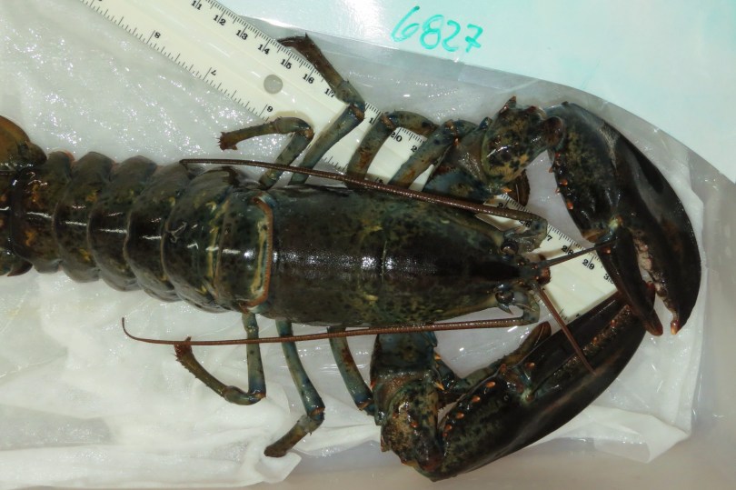
A collaborative paper on lobster shell bacteria has just been published in the journal iScience: “Water temperature and disease alters bacterial diversity and cultivability from American Lobster (Homarus americanus) shells.” This paper investigates what happens to bacterial communities on healthy and sick lobsters as they experience different water temperatures for a year.
I joined this project back in the summer of 2020, towards the end of my first year at UMaine, when I was given a large 16S rRNA gene sequence dataset of bacterial communities from the shells of lobsters. I had been asking around for data as a training opportunity for Grace Lee, who at the time was an undergraduate at Bowdoin College participating in the abruptly cancelled summer Research Experience for Undergrads program at UMaine in summer 2020. Instead, Grace joined my lab as a remote research assistant and we worked through the data analysis over the summer and fall. Grace has since graduated with her Bachelor’s of Science in Neuroscience, obtained a Master’s of Science at Bowdoin, and is currently a researcher at Boston Children’s Hospital while she is applying to medical school.
My first point of contact on the project was Jean MacRae, an Associate Professor of Civil and Environmental Engineering at UMaine, who was the one to lend me the data and who had been working on bacterial community sequencing on other projects which I’ve been involved in. Jean has been involved with MSE, and this is our fourth publication together making her the collaborator at UMaine I have co-authored with the most (although it is a tight race 🙂 ).
Jean introduced me to the original research team, including Debbie Bouchard, who is the Director of the Aquaculture Research Institute and was researching epizootic shell disease in lobsters for her PhD dissertation several years ago; Heather Hamlin, Professor and Director of the School of Marine Sciences; Scarlett Tudor (not pictured), the Education and Outreach Coordinator at the ARI; and Sarah Turner (not pictured), Scientific Research Specialist at ARI. The ARI team is involved in a lot of large-scale aquaculture research, education, and outreach to the industry here in Maine, and the collaborative work I have been doing with them has been a new an engaging avenue of scientific study for me.
In 2022, the research team, along with social science Masters student Joelle Kilchenmann, published a perspective/hypothesis piece which explored unanswered questions about how the movement of microbes, lobsters, and climate could affect the spread of epizootic shell disease in lobsters off the coast of Maine. That perspective paper was a fun exercise in hypothesis generation and asking ‘what if’?
This manuscript is more grounded, and features work that was started in 2016. It examines bacterial communities on the shells of lobsters which were captured off the coast of Southern Maine and maintained in aquarium tanks for over a year. The lobsters were split into three treatment groups: those which were kept in water temperatures that mimicked what they would experience in Southern Maine, colder water to simulated what they would experience in Northern Maine, and hotter water to simulate what they would experience in Southern New England over that year. The original project team wanted to know if temperatures would make a different to their health or microbial communities.
Figure S8. Water temperature regimes, related to STAR Methods. A. Temperatures were obtained through the National Oceanographic Data Center (NODC). NODC temperatures reflect those recorded near Eastport, ME (A); Portland, ME (B); and an average of temperatures from Woods Hole, MA (C) and New Haven, CT (D) was used to represent Southern New England. B. Annual temperature cycles used in this project to represent Southern New England (SNE), Southern Maine (SME) and Northern Maine (NME).
The original project team swabbed lobster shells to obtain bacteria to try and grow in the lab, as well as DNA to sequence and identify whole bacterial communities. Grace and I performed the data analysis to identify which taxa were present in those communities, what happened over time or when the water temperature changed, and what bacteria were present or not in lobsters which died during the study.
Figure S11. Lobster carapace sampling using a sterile cotton swab to obtain bacterial communities from the shell surface, related to STAR Methods. The right side of the dorsolateral area of the cephalothorax was sampled for the baseline sampling, the left side for the Time 1, and the right side again for Time 2.
In addition to wanting to know about temperature, we wanted to know specifically how temperature would affect the bacteria if the lobsters had epizootic shell disease. It is not known what causes epizootic shell disease (which is why it is called ‘epizootic’), but it manifests as pitting in the shells of lobsters. Over time, the pitting can weaken shells and make it difficult for the lobster to molt, or make the lobster susceptible to predators or microbial infections. This type of shell disease had been a huge problem in Southern New England over the past few decades, and in Maine we have seen more cases over time.
Figure S10. Examples of lobster shell disease indices, related to STAR Methods. A) 0, no observable signs of disease, B) 1+, shell disease signs on 1-10% of the shell surface, C) 2+, shell disease signs on 11-50% of the shell surface, D) 3+, shell disease signs on > 50% of the shell surface.
The highlights of this project are here, but you can click the link below to read the entire study and what happened to lobster health and lobster microbes over time.
Shell bacteria from healthy lobsters, often overlooked, were included in the study.
Hotter and colder water temperatures affected shell bacterial communities.
Epizootic shell disease reduced bacterial diversity on lobster shells.
Epizootic shell disease could be induced or exacerbated by the loss of commensal bacteria from shells.
Suzanne L. Ishaq1,2,, Sarah M. Turner2,3, Grace Lee4,5,M. Scarlett Tudor2,3, Jean D. MacRae6, Heather Hamlin2,7, Deborah Bouchard2,3
1 School of Food and Agriculture; University of Maine; Orono, Maine, 04469; USA.
2 Aquaculture Research Institute; University of Maine; Orono, Maine, 04469; USA.
3 Cooperative Extension; University of Maine; Orono, Maine, 04469; USA.
4 Department of Neuroscience, Bowdoin College, Brunswick, ME 04011; USA.
5 Boston Children’s Hospital, Boston, MA 02115; USA.
6 Department of Civil and Environmental Engineering; University of Maine; Orono, Maine, 04469; USA.
7 School of Marine Sciences; University of Maine; Orono, Maine, 04469; USA.
Summary
The American lobster, Homarus americanus, is an economically valuable and ecologically important crustacean along the North Atlantic coast of North America. Populations in southern locations have declined in recent decades due to increasing ocean temperatures and disease, and these circumstances are progressing northward. We monitored 57 adult female lobsters, healthy and shell-diseased, under three seasonal temperature cycles for a year, to track shell bacterial communities using culturing and 16S rRNA gene sequencing, progression of ESD using visual assessment, and antimicrobial activity of hemolymph. The richness of bacterial taxa present, evenness of abundance, and community similarity between lobsters was affected by water temperature at the time of sampling, water temperature over time based on seasonal temperature regimes, shell disease severity, and molt stage. Several bacteria were prevalent on healthy lobster shells but missing or less abundant on diseased shells, although some bacteria were found on all shells regardless of health status.
Over the past few months, a large team of undergraduate, graduate, and postdoctoral researchers, and I, have been processing hundreds of samples from our scallop hatchery microbiome project. As 2022 winds down, so does the first phase of our lab work, and we are taking a well-deserved break over the holidays before we launch additional lab work, data analysis, and manuscript writing in 2023.
In 2021, the Ishaq Lab, collaborators at UMaine, and collaborators at the Downeast Institute ran a pilot project to investigate the bacteria that associate with sea scallop larvae in hatcheries, and how this is develops in relation to bacteria in hatchery tanks over time. For that project, we collected hundreds of culture plates with a specialized media that selects for certain species of bacteria.
When tanks are drained and cleaned every two days, cotton swabs are rolled across part of the bottom or side of the tank and used to inoculate bacteria onto these culture plates. This is part of a routine screening for pathogens, and don’t worry, we aren’t finding bacteria that causes disease in humans. But, these screening plates creates a useful starting point for our research on bacterial community dynamics.
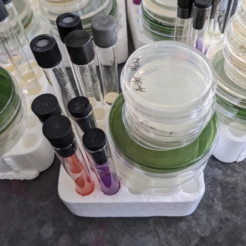
Tank swab samples are used to inoculate TCBS plates to screen for Vibrio and similar bacteria
We received over 200 of these TCBS culture plates, and from them we isolated 140 bacteria in 2021 and early 2022 which we archived at -80 degrees Celsius. This was part of Sarah Hosler’s master’s of science thesis in August of 2022, and has since been passed to Ayodeji Olaniyi for part of his master’s of science thesis.
This fall, we were able to recover 115 of these isolates from the deep freeze, and tested them on 12 different media in duplicate, which created >1800 cultures plates and tubes, and 230 microscope slides!
This massive undertaking would not have been possible without a large team helping with the lab work, including rockstars Ayodeji Olaniyi, Sydney Shair, Keagan Rice, and Lacy Mayo who put in hours and hours leading the efforts on this. We are also grateful to Alaa Rabee, Aaron Williams, Lily Robbins, Ash VanNorwick, and Rebecca Kreeger who provided assistance with media making, inoculating, and the large amount of cleanup (we used glass or autoclavable plastic where possible, and sterilized some single-use plastics to be used as training tools for student education). We were also assisted by Bryanna Dube, who is working on creating outreach/education materials based on our results.
Now, our team will focus on analyzing the results of all these microbiology tests and look for trends. Some will also be heading to the Perry Lab to learn how to perform quantitative polymerase chain reactions (qPCR), in which we use a modified version of DNA replication to count the copies of specific genes. We will use this to look for genes which confirm the identity of our bacteria.
Beginning in summer 2022, the Ishaq Lab has also been part of a state-wide research and commercial collaboration to understand and improve sea scallop production in hatcheries and farms. As part of that project, we received 1500 DNA samples from different hatchery tanks or larvae over the summer/fall rearing season.
Gloria Adjapong is a Postdoctoral Fellow at the UMaine Cooperative Extension Veterinary Diagnostics Lab, and she has been graciously extracting these samples as part of her cross-training in the Ishaq Lab. We will use the extracted DNA to sequence the bacterial communities to identify which bacteria are present, and when, to understand microbial community dynamics over time and in relation to scallop health.
Last week, undergraduate Alexis Kirkendall concluded her 10-week Research Experience for Undergraduates (REU) program at the University of Maine. Over that time, she has been an integral member of the Ishaq lab, and had assisted with lab work for multiple projects, comparing microscopy staining protocols, training students, assisting with laboratory management and safety regulation checks, and more. She picked up skills in animal sample collection, microscopy and staining, parasitology, culture media preparation, DNA extraction, and data visualization in R. Alexis also helped create some of the marketing materials for the Microbes and Social Equity Symposium in July, and facilitated group discussions as a note taker.
At the end of the program, REU students create posters and short presentations of their efforts over the summer. The presentations were last week, but you can check out the poster below.
Alexis is heading back to Ohio for her next year at Heidelberg College where, in addition to studying science, she is leading initiatives to make the campus more accessible and inclusive. But, we hope to see her back in Maine sometime in the future!
This year, the UMaine Student Research Symposium was held in person and virtually, and undergrads and grads from the Ishaq Lab shared their research with the Maine community. You can check out the recorded presentations in the links below.
Pelletier*, E., Taylor, T., Ishaq, S. Abstract 830. Assessing the Veterinary Needs of Rural Maine and Implementing an Effective Management Plan. UMaine Student Symposium (poster presentation). April 15, 2022.
A scientific article led by my colleague Dr. Alaa Rabee at the Desert Research Center in Egypt was just published online and is now available! Dr. Rabee and I have been collaborating remotely on projects related to the bacteria in the rumen of camels, sheep, and cows, as Dr. Rabee’s work focuses on the isolation of bacteria which can degrade plant materials efficiently and could be used to produce biofuels. He will be spending 6 months working in my lab as a visiting scholar, which was delayed until this year because of the pandemic.
Rabee, A.E., Sayed Alahl, A.A., Lamara, M., Ishaq, S.L. 2022. Fibrolytic rumen bacteria of camel and sheep and their applications in the bioconversion of barley straw to soluble sugars for biofuel production. PLoS ONE 17(1): e0262304. Article.
Abstract
Lignocellulosic biomass such as barley straw is a renewable and sustainable alternative to traditional feeds and could be used as bioenergy sources; however, low hydrolysis rate reduces the fermentation efficiency. Understanding the degradation and colonization of barley straw by rumen bacteria is the key step to improve the utilization of barley straw in animal feeding or biofuel production. This study evaluated the hydrolysis of barley straw as a result of the inoculation by rumen fluid of camel and sheep. Ground barley straw was incubated anaerobically with rumen inocula from three fistulated camels (FC) and three fistulated sheep (FR) for a period of 72 h. The source of rumen inoculum did not affect the disappearance of dry matter (DMD), neutral detergent fiber (NDFD). Group FR showed higher production of glucose, xylose, and gas; while higher ethanol production was associated with cellulosic hydrolysates obtained from FC group. The diversity and structure of bacterial communities attached to barley straw was investigated by Illumina Mi-Seq sequencing of V4-V5 region of 16S rRNA genes. The bacterial community was dominated by phylum Firmicutes and Bacteroidetes. The dominant genera were RC9_gut_group, Ruminococcus, Saccharofermentans, Butyrivibrio, Succiniclasticum, Selenomonas, and Streptococcus, indicating the important role of these genera in lignocellulose fermentation in the rumen. Group FR showed higher RC9_gut_group and group FC revealed higher Ruminococcus, Saccharofermentans, and Butyrivibrio. Higher enzymes activities (cellulase and xylanase) were associated with group FC. Thus, bacterial communities in camel and sheep have a great potential to improve the utilization lignocellulosic material in animal feeding and the production of biofuel and enzymes.
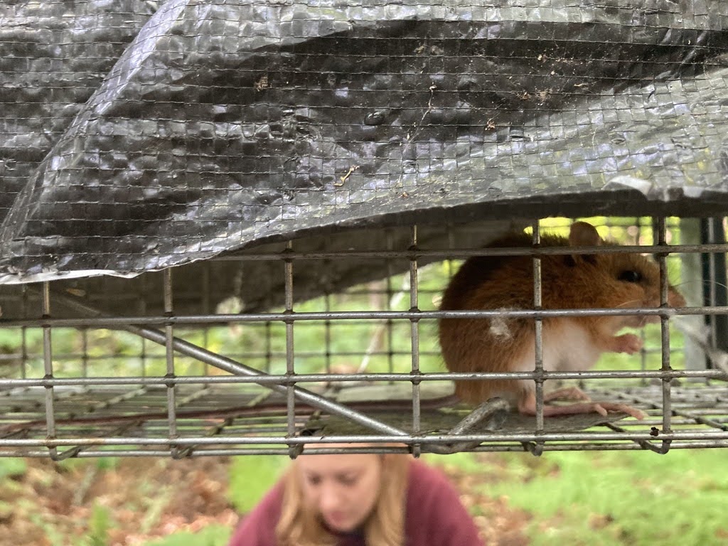
Funded by the University of Maine Rural Health and Wellbeing Grand Challenge Grant Program, this project assesses pathogen carriage by mice and flying squirrels on or near farms in several locations in Maine. We live-capture mice and flying squirrels in traps, collect the poop they’ve left in the trap, and conduct a few other health screening tests in the field before releasing them. To maximize the information we collect while minimizing stress and interference to the animals, information is being collected for other projects in the Levesque Lab at the same time. We will be collecting samples for another few weeks, and then working on the samples we collected in the lab over the fall and winter.
One of the major goals of the funding program, and this project, is to engage students in research. After a few months on the project, some of our students describe their role and their experiences so far…
Marissa Edwards
Undergraduate in Biology
Levesque Lab
Hi! My name is Marissa Edwards and I am an undergraduate research assistant with Danielle Levesque. This summer, my role has been to set traps, handle small mammals, and collect fecal and tissue samples from deer mice.
One of the skills I’ve learned this summer is how to properly ear tag a mouse. To catch mice, we set traps across UMaine’s campus as well as other parts of Maine, including Moosehead Lake, Flagstaff Lake, and Presque Isle.
During our trip to Moosehead Lake, I saw a marten for the first time (it was in one of our traps). I did not know martens existed and initially thought it was a fisher cat. It was both a cool and terrifying experience!
Elise Gudde
Master’s Student of Ecology and Environmental Sciences
Levesque Lab
Hello, my name is Elise Gudde, and I am currently a master’s student at the University of Maine in the Ecology and Environmental Sciences program. I work in Dr. Danielle Levesque’s lab studying small mammal physiology in Maine.
This summer, as a part of the squirrel project, I work to trap small mammal species in Maine, such as white footed mice, deer mice, and flying squirrels in order to determine which species have shifted their range distributions as a result of climate change. Being a part of the research team, this summer has brought me all over Maine! I have been able to travel to Orono, Greenville, New Portland, and Aroostook County to study many interesting mammals. I even got to handle an Eastern chipmunk for the first time! As a member of the animal-handling side of the research team, I also collect fecal and tissue samples from the animals. These samples are then handed off for other members of the team to research in the lab!
Rebecca French
Undergraduate in Animal and Veterinary Sciences
Ishaq Lab
In the beginning of this project, I had no idea what I was getting myself into when I began researching flying squirrels and mice. I came into it with almost no in-person lab experience, so I had a lot to learn.
So far, I have been focusing on making media on petri dishes for culturing bacterial growth and after plating fecal bacteria on said plates; discerning what that growth can be identified as.
We are using media with specific nutrients, and colored dyes, and certain bacteria we are interested in will be able to survive or produce a color change. I have also been performing fecal flotations and viewing possible eggs and parasites under a microscope. What I’ve found most fun about this project is putting into practice what I have learned only in a classroom setting thus far. It is also very satisfying to be a part of every step of the project; from catching mice, to making media, to using that media to yield results and then to be able to have a large cache of information to turn it all into a full fledged project.
Some of the media used to culture bacteria from the feces of mice and squirrels.
Joe Beale
Undergraduate in Animal and Veterinary Sciences
Kamath Lab
Hello! My name is Joseph Beale, and I am an undergraduate at the University of Maine working on the squirrel project as a part of my capstone requirement for graduation. My primary responsibility in this project is the molecular testing of samples obtained from the field. Primarily I will be working with ear punch samples taken from flying squirrels and field mice. DNA extracts from these field samples will be run via qPCR. The results of this qPCR will tell us if these squirrels are carrying any pathogens.
The pathogens we will be testing for are those found in Ixodes ticks. The qPCR panel which we will be running the extracted DNA from the ear punches on tests for Borrelia burgdorferi, the causative agent of Lyme disease, Anaplasma phagocytophilum, the causative agent of anaplasmosis, and Babesia microti, the causative agent of Babesiosis. These pathogens and respective diseases discussed are all transmitted through Ixodes ticks. Deer ticks are the most common and famous of the Ixodes genus. The Ixodes genus encapsulates hard-bodied ticks. Along with deer ticks, Ixodes ticks found in Maine include: woodchuck ticks, squirrel ticks, mouse ticks, seabird ticks, and more. Mice and squirrel are ideal hosts for these Ixodes ticks, therefore becoming prime reservoirs for these diseases. In our research, we are interested in determining the prevalence of these diseases in squirrels and mice as these hosts can spread these diseases to humans and other animals in high tick areas.
qPCR, quantitative polymerase chain reaction, allows for the quantification of amplified DNA in samples. This will help tell us if these pathogens are present in samples and in what capacity. In qPCR provided DNA strands are added to the reaction. These strands match with the genome of the intended pathogens. If the pathogens are present in our samples, the provided DNA strands will bind to the present pathogen DNA. PCR will then work to manufacture billions of copies of this present pathogen DNA.
When not working on this project, I also work in the University of Maine Cooperative Extension Diagnostic Research Laboratory as a part of the Tick Lab. In this position I have honed the molecular biology skills that I will in turn use for the squirrel project.
Hello everyone! My name is Yvonne Booker and I am a rising senior, animal and poultry science major at Tuskegee University in Tuskegee, Alabama. I am interested in animal health research, with a particular focus in veterinary medicine. I’ve always wanted to be a veterinarian, but as I progressed throughout college, I became interested in learning more about animal health and how I might help animals on a much larger and impactful scale–which led me to the REU ANEW program. Currently climate change is causing an increase in global temperatures, putting pressure on animals’ ability to interact and survive within their environment. Consequently, scientists are now attempting to understand not just how to prevent climate change, but how these creatures are adapting to this emerging challenge.
My research experience this summer is geared toward addressing this global issue. I am currently working in Dr. Danielle Levesque’s Lab, which aims to study the evolutionary and ecological physiology of mammals in relation to climate. My project involves conducting a literature review of the microbiome of mammals, to learn more about how their microbial community plays a role in how they adapt in a heat-stressed environment.
Our knowledge of vertebrate-microbe interactions derives partly from research on ectotherms. While this research paves the path for a better understanding of how organisms react to temperature changes, fewer studies have focused on how mammals deal with these extreme temperature shifts—specifically, the abrupt surge in climate change. The ability of endotherms to thermoregulate alters our knowledge of (1) how mammals create heat tolerance against these environmental challenges and (2) how this internal process alters mammals’ adaptability and physiology over time. We suggest that the microbiome plays an essential part in understanding mammals’ heat tolerance and that this microbial community can help researchers further understand the various processes that allow mammals to survive extreme temperatures.
As a student of the REU ANEW program my goal was to go out of my comfort zone and study animals in an applied fashion that would impact animal health on an environmental and ecological scale; and this program was just that! My mentor, Dr. Levesque was wonderful in guiding me through conducting this research, while giving me the independence to create my own voice. The program directors, Dr. Anne Lichtenwalner and Dr. Kristina Cammen, have also been extremely supportive throughout this entire program equipping students with the tools they need to succeed as researchers. Although research was my primary focus this summer, some of my favorite memories involved building community with the students and the staff. From weekly check-ins on zoom to virtual game nights of complete smiles and laughter, this program has been one for the books! The One Health and the Environment approach to this Research Experience for Undergraduate students has encouraged me to build on my curiosity within the field of science, and I’m looking forward to applying what I’ve learned to my career in the future.
Did you know that camels have three stomach chambers or that they have to throw up their own food in order to digest their food properly? Have you felt excluded from science spaces before? Then this blog post is for you!
Allow me to introduce myself.
My name is Myra, and I am a rising senior at SUNY Stony Brook University, where my major is Ecosystems and Human Impact, with a biology minor. In a nutshell, my major is interdisciplinary with a focus on conservation and ecology within human societies.
If I were to describe my college experience in one word I’d pick “surprises”. I never actually saw myself being a scientist in my middle and high school years. I found it hard to care about abstract concepts or theories that felt so far removed from humanity, particularly minority communities. But, during college I found myself falling in love with environmental studies, and along with it, the beautiful complexities that come with being human in our increasingly anthropogenic world.
At UMaine, we focus on the One Health Initiative, which views the health of humans, animals, and the environment as interconnected. When COVID-19 caused everyone to go into lockdown, I was fortunate to find this farm was looking for crew members, with a focus on food security. While certainly not how I planned to spend the summer of 2020, farming for underserved communities is where I saw how impactful One Health was. Organic farmers commonly use plastic mulch as a popular alternative to pesticides for weed suppression. At my home institution, I lead a project on the impacts of microplastics on earthworm health, an Ecotoxicology lab (students of the lab affectionately gave it the nickname “the Worm lab”). We use earthworm health as an indicator of soil health, which in turn is crucial for crop flourishment. The Worm Lab and farming emboldened me to pursue science and, ergo, look for this REU!
At UMaine, I am a member of the Ishaq Lab where I work on the camel metagenome project. Basically, scientists in Egypt raised camels on different diets, then used samples from their feces to sequence their microbial genome. These microbes live in the camel rumen (part of the camel stomach), and help the camel digest their food. What I do with Dr. Ishaq’s lab is, I perform data analysis on these sequences to see how the microbial gene profile changes with different diets. Camels are essential for transportation and food for the communities that rely on them, so finding the most efficient feed for them is important. Camels also release methane depending on their diet so it’s possible humans could control methane production of camels through their diet.
Being a part of the REU ANEW program for 2021 definitely has been an interesting experience, since it is the first time this program has been conducted virtually. Even though I would have loved to have seen everyone in person and spent time in lovely Orono, Maine, I’m glad for the research opportunity as it has further solidified my love of research and the One Health initiative.
Myra’s poster for the REU Research Symposium, virtual, Aug 13, 2021.
A few weeks ago, I sat down with Sheba A-J, one of the producers of the WeTalkScience podcast, to talk about one of my recent publications in the research journal iScience, at which Sheba is also an editor. Listen to find out how lobsters are like humans, how I got involved on a project working with ants and nematodes, and how you can help make science a more welcoming place.
Ishaq, S.L., A. Hotopp, S. Silverbrand, J.E. Dumont, A. Michaud, J. MacRae, S. P. Stock, E. Groden. 2021. Bacterial transfer from Pristionchus entomophagus nematodes to the invasive ant Myrmica rubra and the potential for colony mortality in coastal Maine. iScience 24(6):102663. Article.